CLINICAL CASE
Closed reduction of humeral condylar fracture and elbow luxation in a dog
Reducción cerrada de fractura condilar de húmero y luxación de codo en un canino
Sebastián Cardona R,1 MVZ, Luis-Carlos Muñoz R,1 MVZ, Raúl-Fernando Silva M,1* Ph.D.
1Universidad de Caldas, Facultad de Ciencias Agropecuarias, Grupo de investigación Terapia Regenerativa, A.A Nro. 275, Manizales, Colombia.
*Correspondence: raul.silva@ucaldas.edu.co
Received: August 2014; Accepted: February 2015.
ABSTRACT
Fractures of the distal humerus that involving the condyles often requires extensive surgical approach for treatment, leading to a prolonged recovery time. In a West Highland White Terrier dog two years old, with a fracture of the lateral humeral condyle portion and elbow luxation, was performed a closed technique for luxation reduction, fracture reduction was performed by digital pressure and percutaneous osteosynthesis by introduction of a Kirschner wire of 2 mm diameter, accompanied by Robert-Jones bandage modified for 6 weeks. Limb function was recovered in the immediate postoperative, the wire was removed six weeks after, non-was observed postoperative complications, and full functional recovery of the limb was clinically evident. This suggests that this technique could be an option in cases of condylar fractures in small-sized dogs.
Key words: Dog, fixation, orthopedics (Source: CAB).
Las fracturas de la porción distal del húmero que involucran los cóndilos, frecuentemente exigen para su reparación un amplio abordaje quirúrgico, que conlleva un tiempo de recuperación prolongado. En un canino West Highland White Terrier de dos años de edad, con una fractura de la porción lateral del cóndilo humeral y luxación de codo, se realizó una técnica cerrada de reducción de la luxación, reducción de la fractura mediante presión digital, y osteosíntesis percutánea mediante la introducción de un clavo de kirschner de 2 mm de diámetro, acompañado de vendaje de Robert-Jones modificado durante 6 semanas. El paciente recuperó la función del miembro en el posquirúrgico inmediato, no se evidenciaron complicaciones posquirúrgicas, seis semanas después se retiro el clavo y se evidenció clínicamente recuperación total de la funcionalidad del miembro. Lo anterior sugiere que esta técnica podría ser una opción en casos de fracturas condilares en perros de pequeño porte.
Palabras clave: Fijación, ortopedia, perro (Fuente: CAB).
INTRODUCTION
The humerus fractures make up almost 10% of all appendicular fractures (1). Almost half of the humerus fractures (46.6%), occur in the distal aspect (2). Of these fractures, the humeral condyle is relatively frequent (2).
The reduction of condylar fractures is usually made with wide surgical approaches that allow for the visualization of all structures. These approaches include a portion of the ulnar-humerus of the el joint elbow by osteotomy of the olecranon or by tenotomy of the triceps tendon, among others (3). As a result, the reduction of condyle fractures shows a high incidence in post surgical complications, among which are the reduction of the range of the articular movement and osteoarthritis of the elbow, among others (4).
As a result, the application of biological osteosynthesis concepts (5), through the use of Kirschner wires in a minimally invasive way, can offer great advantages with respect to traditional techniques, such as less postoperative pain, fast healing and less iatrogenic trauma to important structures such as the physes and the articular capsule.
The closed reduction of a lateral traumatic luxation of the humerus-radius-ulnar is described below. The reduction is performed by digital pressure of a fracture of the lateral portion of the condylar humeral and the fixation through the precutaneous application of a Kirschner wire to a canine patient.
CLINICAL CASE
The patient was a West Highland White Terrier dog, adult, two years old and 7.4 kilograms of body mass, that went to consultation after 96 hours of suffering a fall from a height of 3 meters, with severe claudication of the right thoracic limb characterized by lack of support and total absence of articular mobility.
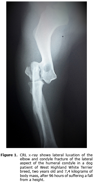
X-rays were made in the medium-lateral and the crown-rump length projections evidencing lateral luxation of the humerus-radius-ulnar articulation and the condylar fracture of the lateral aspect of the humeral condyle (Figure 1).
Because the owner did not have the financial resources to pay for the cost of the treatment through open reduction and internal fixation, and since the owner had to travel quickly to a remote location with no veterinary service available, it was decided to perform an ambulatory treatment. Through traction and counter-traction maneuvers a closed reduction of the luxation was performed and, through the Campbell evaluation method, it was determined that the collateral ligaments were intact (6).
The surgical treatment consisted in a closed reduction of the fracture of the lateral aspect of the humeral condyle by digital pressure. A Kischner wire of 2 mm was introduced by percutaneous access through the lateral fractured aspect, intercondylar until the medial aspect of the humeral condyle, as the only method of fixation (Figure 2). Later, the dog was immobilized with Robert-Jones bandage that was modified during four months.
RESULTS
Limb function was recovered in the immediate postoperative. When the patient recovered his conscience, he started to wander with moderate claudication of the treated limb. No postsurgical complications appeared during the six weeks of clinical support. Four weeks after the removal of the wire, a satisfactory clinical recovery of the functionality of the limb was evidenced. The Robert Jones modified bandage was removed definitively at six weeks. The extension and bending angles of the affected joint were 150 and 40 degrees, and the angles of the contralateral joint were 154 and 30 degrees, respectively; in the x-rays, no compatible signs with osteoarthritis of the joint were found (Figure 3).
DISCUSSION
Although the main objective of the treatment of fractures in the humeral condyles is to restore the join surface and fixation and the stabilization of the condyle to the humeral dyaphisis (7), in cases were an open reduction and internal fixation (RAFI) cannot be performed to allow a fast mobilization of the joint, the surgical treatment is counterproductive (8). In this case, since the RAFI treatment could not be preformed, a closed reduction and external immobilization was performed as an alternative treatment.
Currently, one of the most used techniques is to apply a screw to obtain interfragmentary compression and primary healing of the fracture (7). Although in this case the application of a wire does not produce interfragmentary compression, which could cause the nonoccurrence of joint congruence and even complications such as bad union or non union, in this case, no complications were evidenced during the evaluation period and clinically, during the subjective evaluation, there was an evolution from severe claudication (loss of function) to no claudication. This allows speculating that the performed treatment did not cause it.
Although RAFI is performed to ensure anatomic alignment of the joint surface, a high incidence of complications has been reported, including claudication, osteoarthritis, non-union, osteomyelitis and loss of movement range, among others (9). It is reported that between 28-57% of the dogs that were subjected to a reduction of the fracture of the humeral condyle, had long-term claudication and loss of the function of the affected limb (2).
The traditional method of reduction of fractures known as anatomic reconstruction (10), offers disadvantages with respect to biological osteosynthesis, such as prolonged surgery time, excessive retraction and alteration of the soft surrounding tissue (11). This not only limits the extra osseous blood supply necessary for the formation of the callus (10, 12), but it also reduces the transfer of mononuclear cells; which could not only affect the healing time but could also compromise the resistance to infection and increase the probabilities of bacteria contamination. Unlike the biological osteosynthesis concepts (5), which include the fixation of fractures without altering surrounding soft tissues and the periosteum (12), which among others, the treatment offers advantages such as the preservation of the potential osteogenic of the bruising of the fracture (10, 13), which promotes the osseous regeneration and reduces healing and recovery time (10). In this case, given the impossibility to perform RAFI, the biological osteosynthesis concepts were applied as alternative treatment.
Since no kinetic and/or kinematic methods were available to measure the objective degree of claudication, goniometry was used as an indirect indicator of the degree of joint functionality. This method has been widely used by orthopedics and physiotherapies on humans to quantify the limits of joint movements, determine the degree of loss of function, as a diagnosis aid, to make therapeutic decisions and post treatment control. In canine orthopedist, goniometry has been used to evaluate the efficiency of the treatment in alterations involving joint diseases and it has shown great reliability (14). In this sense, there are reports about the reduction of flexibility and extension degrees of elbow joints in canines subjected to surgery (7). Even though the reported values for flexibility and extension degrees of the elbow range between 20° and 170° respectively (15), the estimated values for this case for elbow joint in flexibility and extension were 40° and 150° in flexibility and extension, but the normal contralateral joint values were of 38° and 154°, respectively. This indicates that the patient recovered a range of mobility close to normal during the evaluation period.
Even though this case presents limitations with respect to diagnostic aids, objective clinical evaluation and the treatment differs from the ideal treatment, in a country with socioeconomic conditions like ours were the ideal economic, technical and logistic conditions for the diagnosis and treatment of certain conditions are not always available, its important to offer therapeutic alternatives different from the natural course of the disease and improve the final result of this evolution, possibly based in body mass and zoo technical attitude of the patient and technical and economical limitations.
The results obtained in this case suggest that the closed reduction of this type of fractures in the procedures described in this clinical case could be considered as an alternative for the treatment of this type of fractures in small dogs.
REFERENCES
1. Johnson JA, Austin C, Breur GJ. Incidence of canine appendicular musculoskeletal disorders in 16 veterinary teaching hospitals from 1980 through 1989. Vet Comp Orthop Traumatol 1994; 7(2):56-69.
2. Bardet JF, Hohn RB, Rudy RL, Olmstead ML. Fractures of the humerus in dogs and cats: a retrospective study of 130 cases. Vet Surg 1983; 12(2):73-77.
3. Simpson A M. Fractures of the humerus. Clin Tech Small Anim Pract 2004; 19(3):120-127.
4. Gordon WJ, Besancon MF, Conzemius MG, Miles KG, Kapatkin AS, Culp WTN. Frequency of post-traumatic osteoarthritis in dogs after repair of a humeral condylar fracture. Vet Comp Orthop Traumatol 2003; 16(1):1-5.
5. Beale, B. Orthopedic clinical techniques femur fracture repair. 2004. Clin Tech Small Anim Pract 2004; 19(3):134-150.
6. Bordelon JT, Reaugh HF, Rochat MC. Traumatic luxations of the apendicular skeleton. Vet Clin Small 2005; 35 (5):1169-1194.
7. Moores A. Humeral condylar fractures and incomplete ossification of the humeral condyle in dogs. In Practice 2006; 28(7):391-397.
8. Schatzer J. The rationale of operative fracture care. Third edition, Springer Berlin Heidelberg New York – USA. 2005; p. 34-35.
9. Kim S E, Hudson C C, & Pozzi A. Percutaneous Pinning for Fracture Repair in Dogs and Cats. 2012. Vet Clin North Am Small Anim Pract 2012; 42(5):963-974.
10. Horstman CL, Beale BS, Conzemius MG, Evans R. Biological Osteosynthesis Versus Traditional Anatomic Reconstruction of 20 Long-Bone Fractures Using an Interlocking Nail: 1994–2001. Vet Surg 2004; 33(3):232-237.
11. McKee W M, Macias C & Innes J F. Bilateral fixation of Y-T humeral condyle fractures via medial and lateral approaches in 29 dogs. J Small Anim Prac 2005; 46(5):217-226.
12. Broos PLO, Sermon A. From unstable internal fixation to biological osteosynthesis. A historical overview of operative fracture treatment. Acta Chir Belg 2004; 104(4):396-400.
13. Kolar P, Schmidt-Bleek K, Schell H, Gaber T, Toben D, Schmidmaier G, Perka C., et al. The early fracture hematoma and its potential role in fracture healing. Tissue Eng Part B Rev 2010; 16(4):427-434.
14. Lamoreaux Hesbach A. Techniques for objective outcome assessment. Clin Tech Small Anim Pract 2007; 22(4):146-154.
15. Millis D & Levine D. Appendix 2. Joint motion and ranges. 2nd edition. En: Canine rehabilitation and physical therapy. Philadelphia, PA: Elsevier. 2014