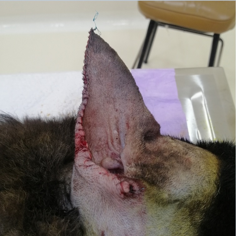
Presencia de mastocitoma en el pabellón auricular en un canino
Presence of mastocytoma in auricular pavilion in a canine

Esta obra está bajo una licencia internacional Creative Commons Atribución-NoComercial-CompartirIgual 4.0.
Mostrar biografía de los autores
Los mastocitos cutáneos y subcutáneos son neoplasias compuestas por mastocitos que forman parte de la piel en los caninos. Es un tumor muy común y el tratamiento va enfocado a la agresividad que pudiera presentar. Ocasionalmente se puede administrar un tratamiento local, sin embargo, este no debe considerarse en pacientes con metástasis. La estadificac ión del tumor es de gran importancia para dar un diagnóstico, tratamiento y pronóstico para aquellos pacientes afectados por esta patología. En este trabajo se presenta el caso clínico de un canino Pastor Alemán de 11 años con la presencia de una estructura ulcerada en el pabellón auricular derecho. Se realizó diagnóstico por citología e histopatológico de mastocitoma canino grado II y baja malignidad de acuerdo con la clasificación de Pakiel. Al paciente se le realizó una resección parcial del pabellón auricular con presencia de bordes limpios en los resultados del estudio histopatológico. El objetivo de este trabajo es reportar un caso clínico con la presencia de mastocitoma en el pabellón auricular en un perro doméstico.
Visitas del artículo 559 | Visitas PDF
Descargas
- Downing S, Chien MB, Kass PH, Moore PE, London CA. Prevalence and importance of internal tandem duplications in exons 11 and 12 of c-kit in mast cell tumors of dogs. Am J Vet Res. 2002; 63(12):1718-23. https://doi.org/10.2460/ajvr.2002.63.1718
- Bellamy E, Berlato D. Canine cutaneous and subcutaneous mast cell tumours: a narrative review. J Small Anim Pract. 2022; 63(7):497-511. https://doi.org/10.1111/jsap.13444
- Oliveira MT, Campos M, Lamego L, Magalhães D, Menezes R, Oliveira R, et al. Canine and feline cutaneous mast cell tumor: A Comprehensive Review of Treatments and Outcomes. Top Companion Anim Med. 2020; 41. https://doi.org/10.1016/j.tcam.2020.100472
- Salvi M, Molinari F, Iussich S, Muscatello LV, Pazzini L, Benali S, et al. Histopathological classification of canine cutaneous round cell tumors using deep learning: A Multi-Center Study. Front Vet Sci. 2021; 26(8):640944. https://doi.org/10.3389/fvets.2021.640944
- Śmiech A, Ślaska B, Łopuszyński W, Jasik A, Bochyńska D, Dąbrowski R. Epidemiological assessment of the risk of canine mast cell tumours based on the Kiupel two-grade malignancy classification. Acta Vet Scand. 2018; 60(1):70. https://doi.org/10.1186/s13028-018-0424-2
- Rinaldi V, Crisi PE, Vignoli M, Pierini A, Terragni R, Cabibbo E, et al. The role of fine needle aspiration of liver and spleen in the staging of low-grade canine cutaneous mast cell tumor. Vet Sci. 2022; 9(9):473. https://doi.org/10.3390/vetsci9090473
- Brown M, Hokamp J, Selmic L E, Kovac R. Utility of spleen and liver cytology in staging of canine mast cell tumors. J Am Anim Hosp Assoc. 2022; 58(4):168–175. https://doi.org/10.5326/JAAHA-MS-7006
- Patnaik AK, Ehler WJ, Macewen EG. Canine cutaneous mast cell tumor: morphologic grading and survival time in 83 dogs. Vet Pathol. 1984; 21(5):469–474. https://doi.org/10.1177/030098588402100503
- de Nardi AB, Dos Santos Horta R, Fonseca-Alves CE, de Paiva FN, Linhares LCM, Firmo BF, et al. Diagnosis, Prognosis and Treatment of Canine Cutaneous and Subcutaneous Mast Cell Tumors. Cells. 2022; 11(4):618. https://doi.org/10.3390/cells11040618
- Berlato D, Bulman-Fleming J, Clifford CA, Garrett L, Intile J, Jones P, et al, Limitations and Recommendations for Grading of Canine Cutaneous Mast Cell Tumors: A Consensus of the Oncology-Pathology Working Group. Vet Pathol. 2021; 58(5):858-863. https://doi.org/10.1177/03009858211009785
- Pratschke KM, Atherton MJ, Sillito JA, Lamm CG. Evaluation of a modified proportional margins approach for surgical resection of mast cell tumors in dogs: 40 cases (2008-2012). J Am Vet Med Assoc. 2013; 243(10):1436-1441. https://doi.org/10.2460/javma.243.10.1436
- Vernker-Van HAJ, Van DGI. Tumors of the external ear. Vet Q. 1998; 20(1):S7. https://doi.org/10.1080/01652176.1998.10807380
- Molino-Díaz VM. Adenocarcinoma de glándulas ceruminosas en un canino: reporte de caso. Rev Med Vet. 2018; (37):95-102. https://doi.org/10.19052/mv.vol1.iss37.11
- Ríos A. Mastocitoma canino y felino. Clín Vet Peq An. 2008; 28(2):35-142. https://ddd.uab.cat/pub/clivetpeqani/11307064v28n2/11307064v28n2p135.pdf
- Álvarez CF, Castro I, Álvarez F. Dermatología en perros y gatos. México: Ed. Jaiser; 2001.
- Chu ML, Hayes GM, Henry JG, Oblak ML. Comparison of lateral surgical margins of up to two centimeters with margins of three centimeters for achieving tumor-free histologic margins following excision of grade I or II cutaneous mast cell tumors in dogs. J Am Vet Med Assoc. 2020; 256(5):567-572. https://doi.org/10.2460/javma.256.5.567


