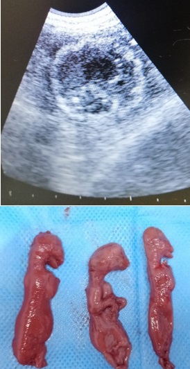
Bioquímica sérica y perfil hematológico de una gata con tres fetos momificados
Serum biochemistry and hematological profile of a cat with three mummified fetuses

Esta obra está bajo una licencia internacional Creative Commons Atribución-NoComercial-CompartirIgual 4.0.
Mostrar biografía de los autores
Los valores bioquímicos y hematológicos del suero se utilizan para
determinar el resultado de enfermedades tanto en animales como en
humanos. En el informe científico presentado, se definieron hallazgos
hematológicos y bioquímicos en la gata, que fue formada como tres
fetos momificados. Una gata de 12 meses - que fue apareado hace
38 días - con quejas de vómitos, anorexia y polidipsia fue llevado al
Centro de Investigación y Aplicación de Animales de la Universidad de
Siirt. Después del examen clínico preliminar, se observó en el examen
ultrasonográfico que el feto no tenía latidos cardíacos y las áreas
hiperecoicas estaban aumentadas. El feto momificado fue
diagnosticado. La momificación fetal es ocasional en las gatas y ha
sido reportada. Se tomaron muestras de sangre para bioquímica
sérica y análisis hematológico. Este informe de caso se presenta
teniendo en cuenta que la bioquímica sérica y los análisis
hematológicos son importantes en casos de fetos momificados en
gatas. Sin embargo, tanto los parámetros hematológicos como
bioquímicos se encontraban dentro de los rangos de referencia. La
ovariohisterectomía se realizó bajo anestesia general. La herida de la
operación estaba completamente curada siete días después de la
cirugía.
Visitas del artículo 377 | Visitas PDF
Descargas
- Johnston SD, Raksil S. Fetal loss in the dog and cat. Vet Clin North Am Small Anim Pract. 1987; 17(3):535-554. https://doi.org/10.1016/S0195-5616(87)50052-3
- Lamm CG, Njaa BL. Clinical approach to abortion, stillbirth, and neonatal death in dogs and cats. Vet Clin North Am Small Anim Pract. 2012; 42(3):501-513. https://doi.org/10.1016/j.cvsm.2012.01.015
- Lefebvre R. Fetal mummification in the major domestic species: current perspectives on causes and management. Vet Med. 2015; 6:233-244. https://www.dovepress.com/getfile.php?fileID=25364
- Gabor LJ, Canfield PJ, Malik R. Haematological and biochemical findings in cats in Australia with lymphosarcoma. Aust Vet J. 2006; 78:456-461. https://doi.org/10.1111/j.1751-0813.2000.tb11856.x
- Yamada T. Serum amyloid A (SAA): A concise review of biology, assay methods and clinical usefulness. Clin Chem Lab Med. 1999; 37:381-388. https://doi.org/10.1515/CCLM.1999.063
- Ceron JJ, Eckersall PD, Martynez-Subiela S. Acute phase proteins in dogs and cats: Current knowledge and future perspectives. Vet Clin Pathol. 2005; 4:85-99. https://doi.org/10.1111/j.1939-165x.2005.tb00019.x
- Wycislo KL, Connolly SL, Slater MR, Makolinski KV. A biochemical survey of free-roaming cats (Felis catus) in New York City was presented to a trap–neuter–return program. J. Feline Med. Surg. 2014; 16(8):657-662. https://doi.org/10.1177/1098612X13517253
- Safak T, Yilmaz O, Ercan K, Yuksel B, Ocal H. A case of vaginal hyperplasia occurred the last trimester of pregnancy in a Kangal bitch. Ankara Univ Vet Fak Derg. 2021; 68(3):307-310. https://doi.org/10.33988/auvfd.764656
- Hossain A, Noor J, Yadav SK. Fetal mummification in a cat. Acta Scientific Vet Sci. 2021; 3(1):19-22. https://actascientific.com/ASVS/pdf/ASVS-03-0120.pdf
- Sabuncu A, Günay Z, Uçmak M, Enginler SÖ, Erzengin ÖM, Kurban I, Kahraman BB. Different sizes and degrees of foetal mummification during pregnancy in a dog: a case report. Inter J. Vet. Sci. 2013; 2(2):75-77. http://www.ijvets.com/pdf-files/Volume-2-no-2-2013/75-77.pdf
- Kajikawa T, Furuta A, Onishi T, Tajima T, Sugii S. Changes in concentrations of serum amyloid A protein, a1-acid glycoprotein, haptoglobin, and C-reactive protein in feline sera due to induced inflammation and surgery. Vet. Immunol. Immunopathol. 1999; 68:91–98. https://doi.org/10.1016/s0165-2427(99)00012-4
- Korman RM, Ceron JJ, Knowles TG, Barker EN, Eckersall PD, Tasker S. Acute phase response to Mycoplasma haemofelis and ‘Candidatus Mycoplasma haemominutum’ infection in FIV-infected and non-FIV-infected cats. Vet. J. 2012; 193:433–438. https://doi.org/10.1016/j.tvjl.2011.12.009
- Tamamoto T, Ohno K, Ohmi A, Seki I, Tsujimoto H. Time-course monitoring of serum amyloid A in a cat with pancreatitis. Vet Clin Pathol. 2009; 38:83–86. https://doi.org/10.1111/j.1939-165X.2008.00082.x
- Javard, R, Grimes C, Bau-Gaudreault L, Dunn M. Acute-phase proteins and iron status in cats with chronic kidney disease. J Vet Intern Med. 2017; 31:457–464. https://doi.org/10.1111/jvim.14661
- Winkel VM, Pavan TL, Wirthl VA, Alves AL, Lucas SR. Serum alpha-1 acid glycoprotein and serum amyloid A concentrations in cats receiving antineoplastic treatment for lymphoma. Am J Vet Res. 2015; 76:983–988. https://doi.org/10.2460/ajvr.76.11.983
- Tamamoto T, Ohno K, Goto-Koshino Y, Tsujimoto H. Serum amyloid A promotes invasion of feline mammary carcinoma cells. J Vet Med Sci. 2014; 76:1183–1188. https://doi.org/10.1292/jvms.14-0108
- Hazuchova K, Held S, Neiger R. Usefulness of acute phase proteins in differentiating between feline infectious peritonitis and other diseases in cats with body cavity effusions. J Feline Med Surg. 2017; 19:809–816. https://doi.org/10.1177/1098612X16658925
- Vilhena H, Figueiredo M, Cerón JJ, Pastor J, Miranda S, Craveiro H, Tvarijonaviciute A. Acute phase proteins and antioxidant responses in queens with pyometra. Theriogenology. 2018; 115:30-37. https://doi.org/10.1016/j.theriogenology.2018.04.010
- Troia R, Gruarin M, Foglia A, Agnoli C, Dondi F, Giunti M. Serum amyloid A in the diagnosis of feline sepsis. J Vet Diagn Investig. 2017; 29:856–859. https://doi.org/10.1177/1040638717722815
- Yuki M, Aoyama R, Nakagawa M, Hirano T, Naitoh E, Kainuma D. A clinical investigation on serum amyloid A concentration in client-owned healthy and diseased cats in a primary care animal hospital. Vet Sci. 2020; 7:1-9. https://doi:10.3390/vetsci7020045
- Kann RK, Seddon JM, Henning J, Meers J. Acute phase proteins in healthy and sick cats. Res Vet Sci. 2012; 93:649–654. https://doi.org/10.1016/j.rvsc.2011.11.007
- Hwang J, Gottdenker N, Min MS, Lee H, Chun MS. Evaluation of biochemical and hematological parameters and prevalence of selected pathogens in feral cats from urban and rural habitats in South Korea. J Feline Med Surg. 2016; 18(6):443-451. https://doi.org/10.1177/1098612X15587572
- Mooney SC, Hayes AA, Matus RE, MacEwen EG. Renal lymphoma in cats: 28 cases (1977-1984). J Am Vet Med. 1987; 191(11):1473-1477. https://pubmed.ncbi.nlm.nih.gov/3693001


