Ultrasonographic determination of anatomical measurements of tendinous structures and palmar metacarpals ligaments in Colombian Creole donkeys
Determinación ultrasonográfica de medidas anatómicas de estructuras tendinosas y ligamentos metacarpianos palmares en burros criollos colombianos


This work is licensed under a Creative Commons Attribution-NonCommercial-ShareAlike 4.0 International License.
Show authors biography
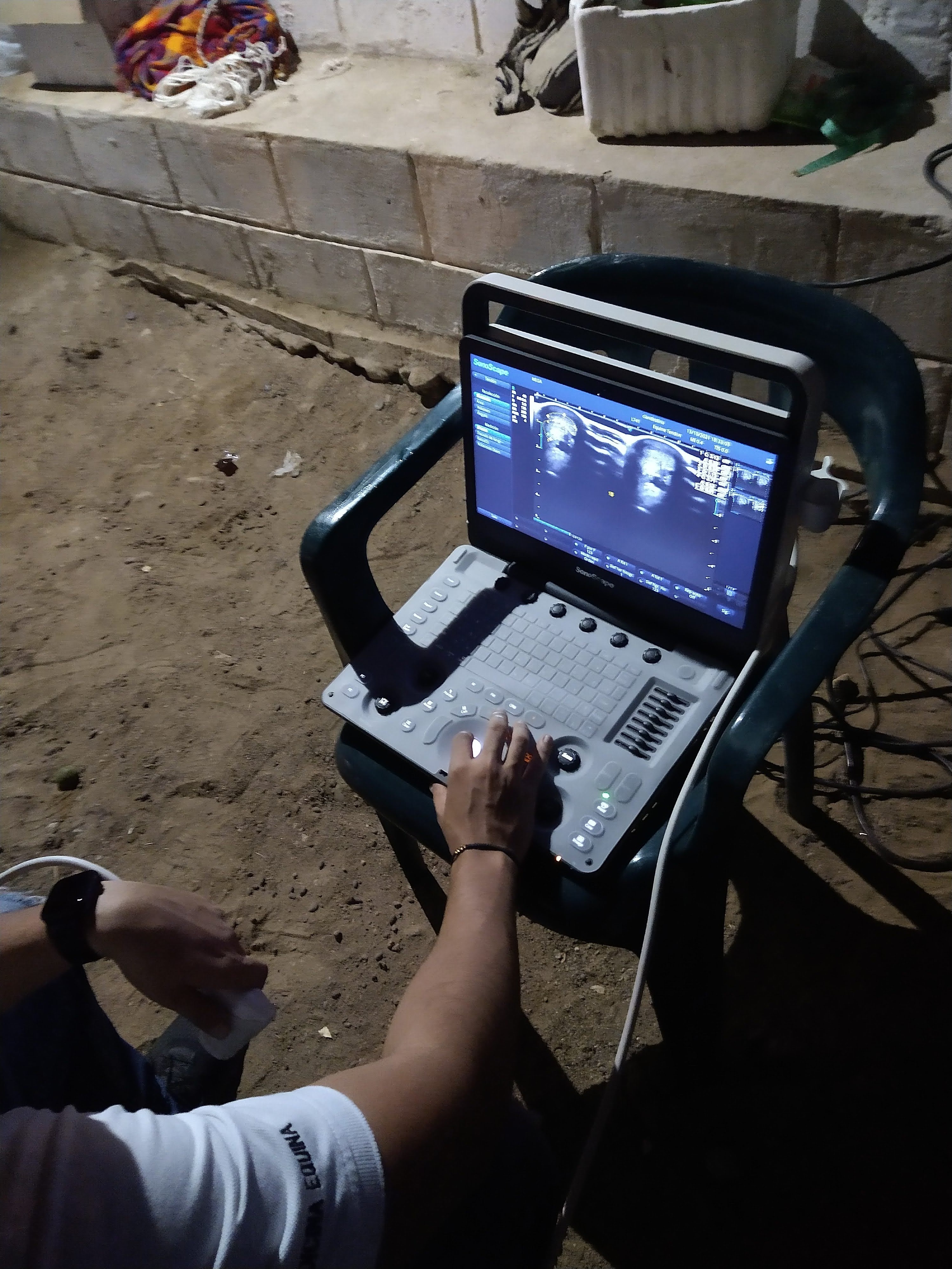
Objective. To provide reference values for anatomical measurements of tendinous structures and metacarpal ligaments in Colombian Creole donkeys using ultrasonography as a measurement tool. Materials and methods. Ultrasonographic examination of the tendons and ligaments of the palmar metacarpal region of limbs was performed in 15 clinically healthy donkeys. The variables to be measured were: cross-sectional area (cm2), lateral medial width (ALM) (cm) and dorsal palmar thickness (EDP) (cm). Results. It was found that there is no difference in the measurements between the two members or in relation to sex. In addition, it was found that the structure with the largest area in the proximal areas (1A and 1B) was the suspensory ligament (0.548 cm2), and in the distal ones (2A and 2B) the deep digital flexor tendon (0.468 cm2). Conclusions. Anatomical measurements of the tendinous structures and the palmar metacarpal ligaments in Colombian Creole donkeys are similar to those found in the international literature. Reference values for anatomical (morphometric) measurements of palmar metacarpal tendons and ligaments in Colombian Creole donkeys were presented.
Article visits 179 | PDF visits
Downloads
- Buthen F, Thieman A. Donkeys are different. J. Equine Vet. Sci. 2015; 35:376-382. http://dx.doi.org/10.1016/j.jevs.2015.03.005
- Kiros A, Gezahegn M, Aylate A. A Cross Sectional Study on Risk Factors Associated with Lameness of Working Donkeys in and around Hawassa, Ethiopia. J. Anim. Health Prod. 2016; 4(3):87-94. http://dx.doi.org/10.14737/journal.jahp/2015/4.3.87.94
- Carluccio A, Noto F, Parrillo S, Contri A, De Amicis I, Gloria A et al. Transrectal ultrasonographic evaluation of combined utero-placental thickness during the last half of pregnancy in Martina Franca donkeys. Theriogenology. 2016; 86(9):2296-2301. https://doi.org/10.1016/j.theriogenology.2016.07.025
- Laus F, Paggi E, Marchegiani A, Cerquetella M, Spaziante D, Faillace V et al. Ultrasonographic biometry of the eyes of healthy adult donkeys. Vet. Rec. 2014; 174(13):326. https://doi.org/10.1136/vr.101436
- Nocera I, Aliboni B, Sgorbini N, Gracia L, Conte G, Bren L et al. Ultrasonographic Appearance of Elbow Joints in a Population of Amiata Donkeys. J Equine Vet. Sci. 2020; 94:103242. https://doi.org/10.1016/j.jevs.2020.103242.
- Rantanen NW. The use of diagnostic ultrasound in limb disorders of the horse: a preliminary report. J Equine Vet. Sci. 1982; 2(2):62–64. https://doi.org/10.1016/ S0737-0806(82)80021-X
- Nazem M, Sajjadian S, Vosough D, Mirzaesmaeili A. Topographic Description of Metacarpal Tendons and Ligaments of Anatoly Donkey by Ultrasonography and Introducing a New Ligament. Anat. Sci. J. 2015; 12(4):153-160. http://anatomyjournal.ir/article-1-119-en.html
- Salem M, El-Shafaey E, Mosbah E, ZaghloulA. Ultrasonographic, Computed Tomographic, and Magnetic Resonance Imaging of the Normal Donkeys (Equus asinus) Digit. J Equine Vet. Sci. 2019;74:68-83. https://doi.org/10.1016/j.jevs.2018.12.019
- Herrera Y, Rugeles C, Ramírez C. Perfil energético, proteico y mineral de burros criollos (Equus asinus) colombianos. Arch. Zootec. 2018; 67(260):512-516. https://doi.org/10.21071/az.v0i0.3881
- Herrera Y, Rugeles C, Vergara O. Perfil hematológico del burro criollo (Equus asinus) colombiano. Rev Colombiana Cienc Anim. 2017; 9(2):158-163. https://doi.org/10.24188/recia.v9.n2.2017.553.
- Cardona J, Reyes B, Martínez M. Cronometría dentaria en equinos. Primera edición. Fondo editorial Universidad de Córdoba. 2019. https://repositorio.unicordoba.edu.co/ handle/ucordoba/2204.
- Burden F. Practical feeding and condition scoring for donkeys
- and mules. Equine vet educ. 2011;24(11):589-596.
- https://doi.org/10.1111/j.2042-3292.2011.00314.x
- Boehart S, Arndt G, Carstanjen B. Ultrasonographic morphometric measurements of digital flexor tendons and ligaments of the palmar metacarpal region in haflinger horses. Anat. Histol. Embryol. 2010; 39(4):366–375. https://doi. org/10.1111/j.1439-0264.2010.01003.x
- Reyes-Bossa B, Medina-Ríos H, Cardona-Álvarez JA. Evaluación de medidas morfométricas de tendones y ligamentos metacarpales palmares por ultrasonografía en caballos criollos colombianos. Rev MVZ Cordoba. 2020; 25(2):e1863. https://doi.org/10.21897/rmvz.1863.
- Pickersgill C, Marr C, Reid S. Repeatability of diagnostic ultrasonography in the assessment of the equine superficial digital flexor tendon. Equine Vet J. 2001; 33(1):33-37. https:// doi.org/10.2746/042516401776767494
- Donkey santuary. The clinical companion of the donkey: second edition. Troubador publishing; 2018. https://www.thedonkeysanctuary.org.uk/what-we-do/for-professionals/resources/clinical-companion
- Nazem N, Sajjadian S. Anatomic assessment of tendons and ligaments of palmar surface of metacarpus in Anatoly donkey and its comparison with horse. J. Vet. Res. 2015; 70(4):424-419 https://dx.doi.org/10.22059/jvr.2016.56462
- Nazem N, Sajjadian. Anatomical transverse magnetic resonance imaging study of ligaments in palmar surface of metacarpus in Miniature donkey: identification of a new ligament. Folia Morphol. 2017; 76(1):110–116. https://dx.doi.org/10.5603/FM.a2016.0032


