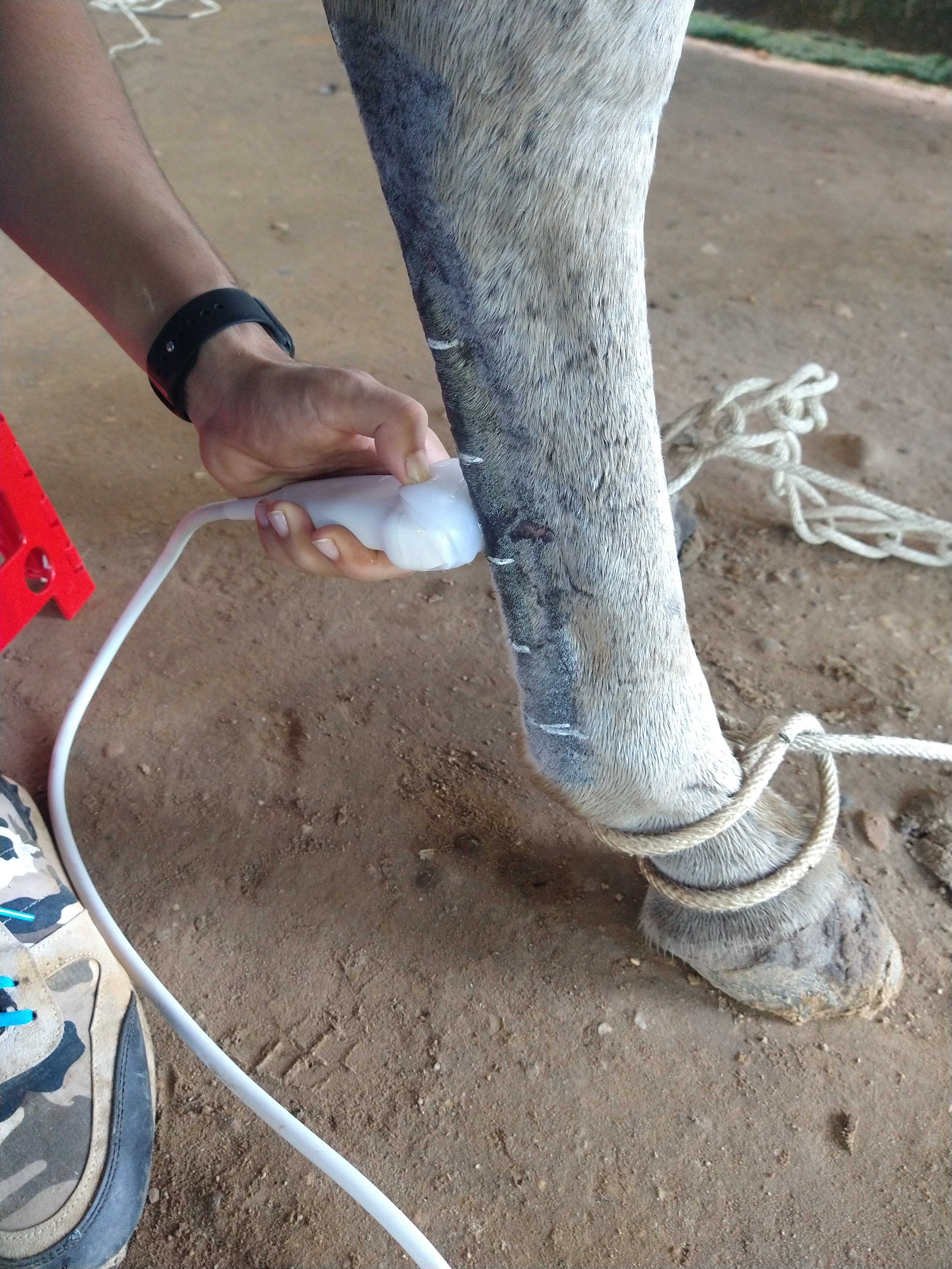
Ultrasonographic evaluation of morphometric measurements of plantar metatarsal tendons and ligaments in Colombian Creole horses
Evaluación ultrasonográfica de medidas morfométricas de tendones y ligamentos metatarsianos plantares en caballos criollos colombianos


This work is licensed under a Creative Commons Attribution-NonCommercial-ShareAlike 4.0 International License.
Show authors biography
Objective. The objective of the present study was to determine the morphometric values of the plantar metatarsal tendons and ligaments in clinically healthy animals. Materials and methods. Thirty animals were used throughout the study, the plantar metatarsal tendons and ligaments were evaluated starting from the plantaromedial aspect of the proximal region to the insertion of the suspensory branches in the sesamoids bones. The variables to be studied in each structure were cross-sectional area (cm2), lateral medial width (ALM) (cm) and dorsal palmar thickness (EDP) (cm). Results. It was found that the structure with the largest area in the proximal regions was the suspensory ligament (0.858 cm2) followed by the lateral digital flexor (0.759 cm2), in regions 1B and 2A the largest structure remained the suspensory ligament and in the region 2B, the deep digital flexor tendon was the largest structure (0.804 cm2). Conclusions. The behavior of the variables in the Colombian Creole Horse is similar to that reported in the literature and finally the reference values of the morphometric measurements of plantar metatarsal tendons and ligaments in this breed are presented.
Article visits 286 | PDF visits
Downloads
- Decon L, Reef V, Leduc L, Navas c. Pocket-Sized Ultrasound Versus Traditional Ultrasoun Images in Equine Imaging: A Pictorial Essay. Journal Eq Vet Sci. 2021; 104:103672. https://doi.org/10.1016/j.jevs.2021.103672
- Bastiani G, De La Côrte F, Brass K, Cantarelli C, Malfestio L, Schwingel D, Silva T, et al. Ultrasonographic, macroscopic and histological characterization of the proximal insertion of the suspensory ligament in Crioulo horses. Pesq Vet Bras. 2019; 39(5):355-363 https://doi.org/10.1590/1678-5150-PVB-5854
- Vandenberghe A, Broeckx S, Beerts C, Seys B, Zimmerman M, Verweire I, et al. Tenogenically Induced allogenic Mesenchymall stem cells for the treatment of proximal suspensory ligamet desmitis in a horse. Front Vet Sci. 2015; 10(2):49. https://doi.org/10.3389/fvets.2015.00049
- Carnicer D, Coudry V, Denoix JM. Ultrasonographic examination of the palmar aspect of the pastern of the horse: sesamoidean ligaments. Equine Vet Educ. 2012; 25(5):256-263. http://dx.doi.org/10.1111/j.2042-3292.2012.00383.x
- Gallego R, Monsalve J, Ospina D, Leysner J. Determinación de lesiones y signos clínicos en caballos criollos colombianos sometidos a cabalgata. FAGROPEC. 2018; 10(1):45-48. https://www.uniamazonia.edu.co/revistas/index.php/fagropec/article/view/1548
- Reyes B, Medina H, Cardona J. Evaluación de medidas morfométricas de tendones y ligamentos metacarpales palmares por ultrasonografía en caballos criollos colombianos. Rev MVZ Córdoba. 2020; 25(2):e1863. https://doi.org/10.21897/rmvz.1863
- Cardona J, Reyes B, Martínez M. Cronometría dentaria en equinos. Primera edición. Fondo editorial Universidad de Córdoba: Colombia; 2019. https://repositorio.unicordoba.edu.co/ handle/ucordoba/2204
- Cardona J, Reyes B, Martínez M. Semiología y propedéutica clínica del aparato locomotor en grandes animales. Primera edición. Fondo editorial Universidad de Córdoba: Colombia; 2019. https://repositorio.unicordoba.edu. co/handle/ucordoba/2203
- Nazem M, Sajjadian S, Vosough D, Mirzaesmaeili A. Topographic Description of Metacarpal Tendons and Ligaments of Anatoly Donkey by Ultrasonography and Introducing a New Ligament. ASJ 2015;12(4):153-160. http://anatomyjournal.ir/browse.php?a_code=A-10-104-3&slc_lang=en&sid=1
- Denoix J, Farres D. Ultrasonographic imaging of the proximal third interosseous muscle in the pelvic limb using a plantaromedial approach. J Equine Vet Sci. 1995; 15:346-350. https://doi.org/10.1016/S0737-0806(07)80544-2
- Pickersgill C, Marr C, Reid S. Repeatability of diagnostic ultrasonography in the assessment of the equine superficial digital flexor tendon. Equine Vet J. 2001; 33(1):33-37. https://doi.org/10.2746/042516401776767494
- Costello J, Kent A, Wakas A, Fugslang L, Harrison A. The Equine Hindlimb Proximal Suspensory Ligament: an Assessment of Health and Function by Means of Its Damping Harmonic Oscillator Properties, Measured Using an Acoustic Myography System: a New Modality Study. Journal Eq Vet Sci. 2018; 71:21-26. https://doi.org/10.1016/j.jevs.2018.09.006
- Rabba S, Petrucci V, Petrizzi L, Giommi D, Busoni V. B-Mode Ultrasonographic Abnormalities and Power Doppler Signal in Suspensory Ligament Branches of Nonlame Working Quarter Horses. J Equine Vet Sci. 2020;94: 103254. https://doi.org/10.1016/j.jevs.2020.103254


