Demographic factors and clinical features regarding dogs with calcium oxalate urolithiasis in Mexico
Factores demográficos y características clínicas en perros con urolitiasis de oxalato de calcio en México


This work is licensed under a Creative Commons Attribution-NonCommercial-ShareAlike 4.0 International License.
Show authors biography
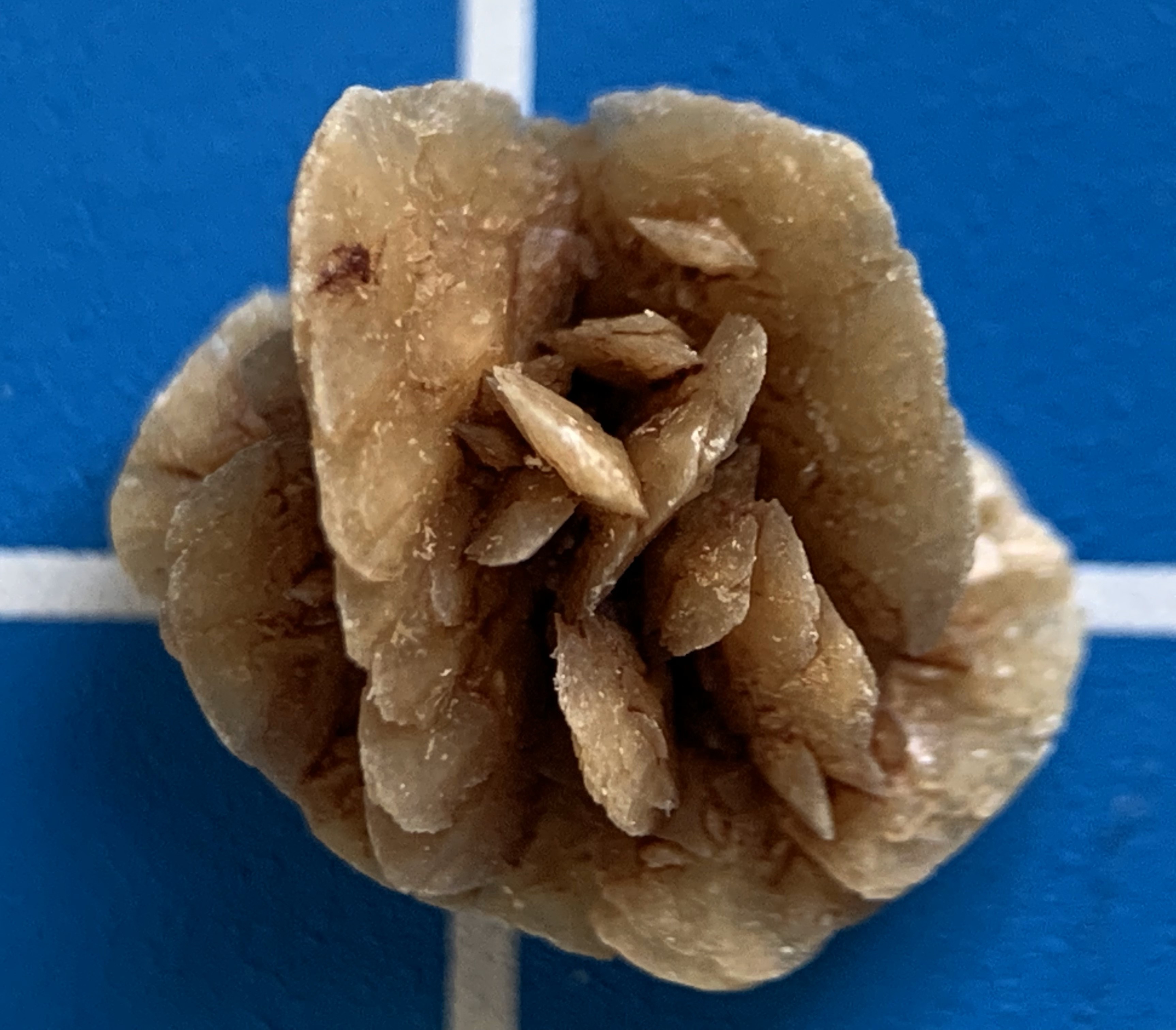
Objective. To identify the most frequent demographic features and clinical findings in dogs with calcium oxalate urolithiasis (CaOx) in Mexico. Materials and methods. Dogs with CaOx uroliths were selected from the laboratory database, and a comparison analysis was performed between two different dog populations to identify the demographic risk factors. Clinical data for urinalysis and the radiographic and physical characteristics of the uroliths were described. The statistical analysis was descriptive, and association measures and multivariate logistic regression were used to identify risk factors. Results. In Mexico, CaOx uroliths accounted for 27.3% of the cases, making it the second most common urolithiasis in dogs. Males, small-sized dogs, and dogs older than 6 years had higher probabilities (p<0.001) of developing CaOx urolithiasis. The clinical findings observed in dogs with this urolithiasis were a mean urine pH of 6.5, urine specific gravity greater than 1.030, highly radiopaque uroliths, multiple and shorter than 20 mm in length. Overall, 31.8% of the CaOx uroliths came from urolithiasis recurring dogs. Conclusions. The identification of the epidemiological features of dogs with CaOx urolithiasis in Mexico, such as sex, breed, size, and age, as well as urinalysis and radiographic findings, may contribute to predict the composition of CaOx uroliths before their extraction and earlier identification in predisposed dogs.
Article visits 426 | PDF visits
Downloads
- Mendoza-López CI, Del-Angel-Caraza J, Quijano-Hernández IA, Barbosa-Mireles MA. Analysis of lower urinary tract disease of dogs. Pesqui Veterinária Bras. 2017;37:1275–1280. https://doi.org/10.1590/s0100-736x2017001100013
- Bende B, Kovacs KB, Solymosi N, Nemeth T. Characteristics of urolithiasis in the dog population of Hungary from 2001 to 2012. Acta Vet Hung. 2015; 63:323–336. https:// doi.org/10.1556/004.2015.030
- Hesse A, Neiger R. A colour Handbook of Urinary stones in small animal medicine. Bonn, Germany: Manson Publishing;2009
- Houston D, Weese H, Vanstone N, Moore A, Weese J. Analysis of canine urolith submissions to the Canadian Veterinary Urolith Centre, 1998–2014. Can Vet J la Rev. 2017; 58:45–50. htts://www.ncbi.nlm.nih.gov/pmc/articles/PMC5157737/
- Lulich JP, Osborne CA, Albasan H, Koehler LA, Ulrich LM, Lekcharoensuk C. Recent shifts in the global proportions of canine uroliths. Vet Rec .2013; 172(4):363. https://doi.org/10.1136/vr.101056
- Blavier A, Sulter A, Bogey A, Novelli K , Billiema z B . Re sul t s o f in f ra red spectrophotometry analysis of 1131 canine urinary stones , collected in France from 2007 to 2010 . Prat médicale Chir l’animal Cie. 2012; 47:7–16. https://doi.org/10.1016/j.anicom.2011.11.001
- Brandenberger-Schenk F, Rothenanger E, Reusch CE, Gerber B. Uroliths of dogs in Switzerland from 2003 to 2009. Schweiz. Arch. Tierheilkd. 2015; 157: 41–48. https://doi.org/10.17236/sat00003
- Vrabelova D, Silvestrini P, Ciudad J, Gimenez JC, Ballesteros M, Puig P, et al. Analysis of 2735 canine uroliths in Spain and Portugal. A retrospective study: 2004-2006. Res Vet Sci. 2011; 91:208–211. https://doi.org/10.1016/j.rvsc.2010.12.006
- Hesse A , Orzekowsky H , Neiger R . Originalarbeit Urolithiasis beim Hund – 15494 Analyse ergebnisse und anam estische Daten aus dem Zeitraum 1979 – 2007 Originalarbeit Kleintierpraxis. 2012; 57:633–639. https://doi.org/10.2377/0023-2076-57-633
- Roe K, Pratt A, Lulich JP, Osborne CA, Syme HM. Analysis of 14,008 uroliths from dogs in the UK over a 10-year period. J Small Anim Pract. 2012; 53:634–640. https://doi. org/10.1111/j.1748-5827.2012.01275.x
- Mendoza-López CI, Del-Angel-Caraza J, AkéChiñas MA, Quijano-Hernández IA, BarbosaMireles MA. Epidemiology of urolithiasis in dogs from Guadalajara City, Mexico. Vet Mex OA. 2019; 6(1):1-14. https://doi:10.22201/fmvz.24486760e.2019.1.585
- D e Lim a Sil va C , Ci n t ra Al ve s C , Meirelles Wilkes Burton A, Crivellenti Borin S, Mariani O, Honsho Kan D, et al. Sensitivity of urolithiasis detection using urinary, radiography and ultrasound parameters. Semin Agrar. 2017; 38:3599–3604. https://doi.org/10.5433/1679-0359.2017v38n6p3599
- Bartges JW, Callens AJ. Urolithiasis. VetClin North Am Small Anim Pract. 2015;45(4):747-768. https://doi:10.1016/j.cvsm.2015.03.001
- Gnanandarajah JS, Abrahante JE, Lulich JP, Murtaugh MP. Presence of Oxalobacter formigenes in the intestinal tract is ssociated with the absence of calcium oxalate urolith formation in dogs. Urol Res. 2012; 40(5):467-473. https://doi:10.1007/s00240-011-0451-1
- Okafor CC, Lefebvre SL, Pearl DL., Yang M, Wang M, Blois SL, et al. Risk factorsassociated with calcium oxalate urolithiasis in dogs evaluated at general care veterinaryhospitals in the United States. Prev. Vet. Med. 2014; 115:217–228. https://doi.org/10.1016/j.prevetmed.2014.04.006
- Kennedy SM, Lulich JP, Ritt MG, Furrow E. Comparison of body condition score and urinalysis variables between dogs with and without calcium oxalate uroliths. J Am Vet Med Assoc. 2016; 249:1274–1280. https://doi.org/10.2460/javma.249.11.1274.
- Wisener LV,Pearl DL, Houston DM, Reid-Smith RJ, Moore AEP. Risk factors for the incidence of calcium oxalate uroliths or magnesium ammonium phosphate uroliths for dogs in Ontario, Canada from 1998 to 2006. Prev Vet Med. 2010; 95:144-151. https://doi.org/10.1016/j.prevetmed.2010.02.016
- Pastor N, Caballé NC, Santella Massimo, E zqu e r ra L J ,Ta ra zon a R , Du ran E . E p idemiologicalst udyofaninemammary tumors:age,breed,size andmalignancy. Austral J Vet. 2018; 50:143-147.http://dx.doi.org/10.4067/S071981322018000300143
- Hunprasit V, Osborne CA, Schreiner PJ, Bender B, Lulich JP. Epidemiologic evaluation of canine urolithiasis in Thailand from 2009 to 2015. Res Vet Sci. 2017; 115:366–370. https://doi.org/10.1016/j.rvsc.2017.07.008
- Hesse A, Frick M, Orzekowsky H, Failing K, Neiger R. Canine calcium oxalate urolithiasis: Frequency of Whewellite and Weddellite stones from 1979 to 2015. Can Vet J. 2018; 59(12):1305-1310. https://www.ncbi.nlm.nih.gov/pmc/articles/PMC6237259/
- Hunprasit V, Schreiner PJ, Bender JB, Lulich JP. Epidemiologic evaluation of calcium oxalate urolithiasis in dogs in the United States: 2010-2015. J Vet Intern Med. 2019; 33(5):2090-2095. https://doi.org/10.1111/jvim.15613
- Aké-Chiñas MA, Mendoza-López CI, DelAngel-Caraza J, Quijano-Hernández IA, Rodríguez-Alarcón CA, Barbosa-Mireles MA. Urolitiasis de estruvita en perros: Características epidemiológicas y clínicas en México. Revista MVZ Córdoba. 2021;27(1):e2338. https://doi.org/10.21897/rmvz.2338
- Turudic D, Batinic D, Golubic AT, Lovric M, Milosevic D. Calcium oxalate urolithiasis in children: urinary promoters/inhibitors and role of their ratios. Eur J Pediatr. 2016; 175(12):1959-1965. https://doi. org/10.1007/s00431-016-2792-9
- Lulich JP, Berent AC, Adams LG, Westropp JL, Bartges JW, Osborne CA. ACVIM Small Animal Consensus Recommendations on the Treatment and Prevention of Uroliths in Dogs and Cats. J Vet Intern Med. 2016; 30:1564–1574. https://doi.org/10.1111/jvim.14559
- Adams LG, Williams JC Jr, McAteer JA, Hatt EK, Lingeman JE, Osborne CA. In vitro evaluation of canine and feline calcium oxalate urolith fragility via shock wave lithotripsy. Am J Vet Res. 2005; 66(9):1651-1654.https://doi:10.2460/ajvr.2005.66.1651
- Koehler LA, Osborne CA, Buettner MT, Lulich JP. Canine Uroliths : Frequently Asked Questions and Their Answers. Vet Clin North Am Small Anim Pract. 2008; 39:161-181. https://doi.org/10.1016/j. cvsm.2008.09.007
- Ogawa N, Sato S, Ida K, Kato K, Ariyoshi Y, Wada K, Nasu Y, Kanazawa S. Evaluation o f U rina ry S tone Compo si tion and Differentiation between Urinary Stonesand Phleboliths Using Single-source Dualenergy Computed Tomography. Acta Med Okayama. 2017; 71(2):91-96. https://doi.org/10.18926/amo/54976
Information


