Gossypiboma secondary to ovariohysterectomy in a dog
Gossypiboma secundario a ovariohisterectomía en una perra


This work is licensed under a Creative Commons Attribution-NonCommercial-ShareAlike 4.0 International License.
 Perfil Google Scholar
Show authors biography
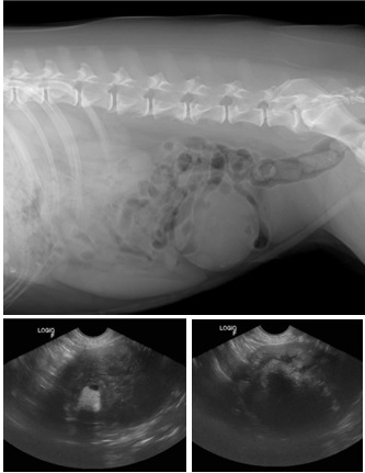
In veterinary medicine, intra-abdominal foreign bodies are a possible secondary complication of surgical procedures. Gossypiboma is defined as a forgotten textile material after surgery and is considered rare in veterinary medicine because most cases go unreported. The objective of this work is to report the diagnosis of an intra-abdominal gossypiboma in a dog, secondary to theovaryhysterectomy procedure. A 10-year-old Labrador dog had been sterilized about eight years ago. During the clinical analysis, a firm-looking increase in volume was found in the right mesogastric region, without the presence of pain on palpation. For further investigation, the patient was referred for additional imaging tests, such as x-ray and ultrasound. During inspection of the abdominal cavity, a firm circular mass was observed adhered to the omentum and jejunum, caudal to the right kidney. Resection of the mass was performed, and then the mass sample was sent for histopathological examination, which revealed an encapsulated foreign body, consistent with surgical gauze. Gossypiboma is one of the possible complications in surgical procedures in the abdominal cavity, but it is rarely reported in the veterinary literature.
Article visits 32 | PDF visits
Downloads
- Carvalho JB, Vinhaes JC. Corpo estranho retido na cavidade abdominal durante onze anos. Rev Col Bras Cir. 2004; 31(1):68-70. http://dx.doi.org/10.1590/S0100-69912004000100013
- Marques LM, Carlos RSA, Silva EB, Clark RMO, Sampaio KMOR, Harvey TV. Imperícia e negligência em ovariosalpingohisterectomia de uma cadela - relato de caso. Rev Bras Med Vet. 2014; 36(4):425-429. https://bjvm.org.br/BJVM/article/view/551/425
- Forster K, Anderson D, Yool DA, Wright C, Burrow R. Retained surgical swabs in 13 dogs. Vet Rec. 2011; 169(13):337. https://doi.org/10.1136/vetreccr.d4396rep
- Chagas NFA, Agnollitto PM, Mauad FM, Barreto ARF, Muglia VF, Junior EJ. Avaliação por imagem dos gossipibomas abdominais. Radiol Bras. 2012; 45(1):53-58 https://doi.org/10.1590/S0100-39842012000100012
- Rodriguez FR, Kirby BM, Ryan J. Evaluation of factors associated with retainedsurgical sponge in veterinary patients: a survey of veterinary practitioners. J Small Anim Pract. 2018; 59:570–577. https://doi.org/10.1111/jsap.12873
- Oliveira R, Matsui A, Ribeiro JO, Simionato G, Simamura AC, Canola J, Camplesi A, Vasconcelos RO, Moraes PC. Clinical and pathological aspects of gossypiboma in a dog: case report. Arq Bras Med Vet Zootec. 2019; 71(1):102–108. https://doi.org/10.1590/1678-4162-10320
- Martins MCB, Amaral RPG, Andrade CS, Lucato TL, Leite AC. Características de imagem na ressonância magnética de gossipiboma intracraniano: relato de caso e revisão da literatura. Radiol Bras. 2009; 42(6):407-409. https://doi.org/10.1590/S0100-39842009000600016
- Day JL, Pechman RD, Bahr RJ. Migration of a retained surgical swab into the jejunum in a dog. J Small Anim Pract. 2012; 53(12):705-708. https://doi.org/10.1111/j.1748-5827.2012.01286.x
- Deschamps JY, Roux FA. Extravesical textiloma (gossypiboma) mimicking a bladder tumor in a dog. J Am Anim Hosp Assoc. 2009; 45(2):89-92. https://doi.org/10.5326/0450089
- Merlo M, Lamb CR. Radiographic and ultrasonographic features of retained surgical sponge in eight dogs. Vet Radiol Ultrasound. 2000; 41(3):279–283. https://doi.org/10.1111/j.1740-8261.2000.tb01491.x
- Mai W, Ledieu D, Venturini L, Fournel C, Fau D, Palazzi X, Magnol JP. Ultrasonographic appearance of intra-abdominal granuloma secondary to retained surgical sponge. Vet Radiol Ultrasound. 2001; 42(2):157-160. https://doi.org/10.1111/j.1740-8261.2001.tb00919.x
- Singh R, Mathur RK, Patidar S, Tapkire R. Gossypiboma: its laparoscopic diagnosis and removal. Surg Laparosc Endosc Percutan Tech. 2004; 14(5):304-305. https://doi.org/10.1097/00129689-200410000-00017
- Iglessias AC, Salomão RM. Intra-abdominal gossypiboma: study of 15 cases. Rev Col Bras Cir. 2007; 34(2):105-113. https://doi.org/10.1590/S0100-69912007000200008
Information


