Radius and ulna locking plate osteosynthesis in giant anteater (Myrmecophaga tridactyla)
Osteosíntesis de radio y ulna con placa bloqueada en oso hormiguero gigante (Myrmecophaga tridactyla)

This work is licensed under a Creative Commons Attribution-NonCommercial-ShareAlike 4.0 International License.
Show authors biography
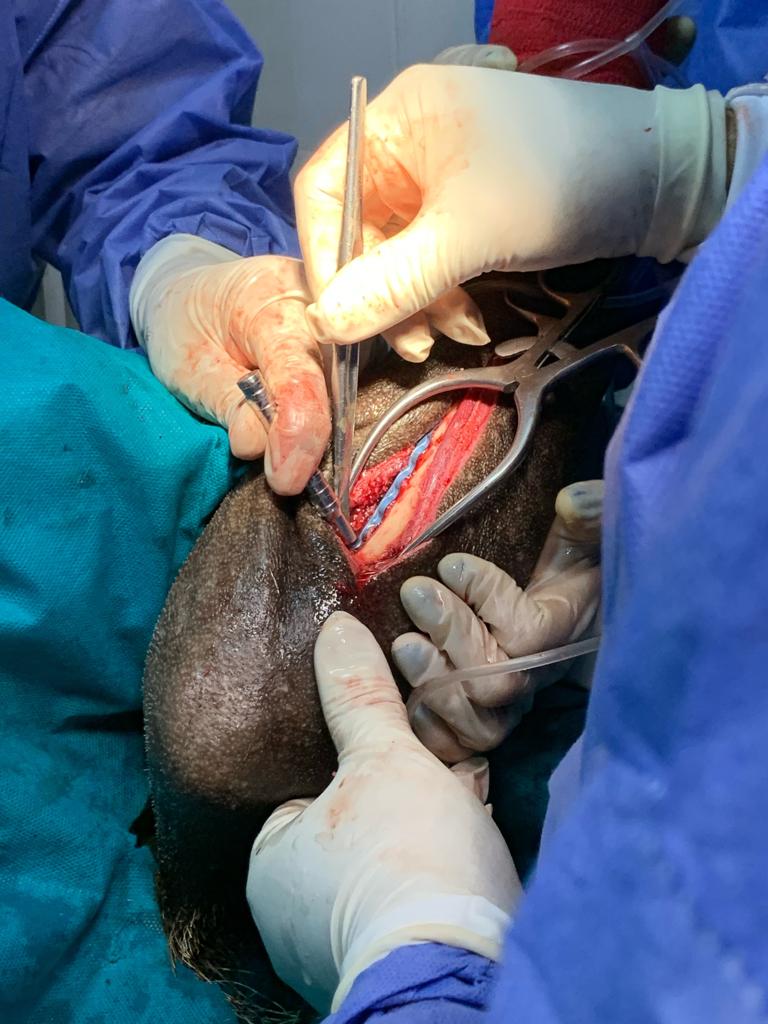
Osteosynthesis is the surgical technique performed in bone fractures for the reduction and stabilization of the bone fragments in a suitable position, allowing the patient to recover their motor activity. This report deals with the case of a Giant Anteater in the municipality of Yopal, Colombia, which was admitted to the veterinary service due to a thoracic limb injury caused by a car accident. The treatment instituted was surgical after diagnosing a fracture of the radio-ulnar diaphysis. Resolution is addressed through osteosynthesis of both bone structures with a double locked titanium plate system under an anesthetic plan of ketamine 4 mg/Kg, dexmedetomidine 0.025 mg/kg and midazolam 0.1 mg/Kg; Maintenance was performed with isoflurane at a rate of 3% as the beginning of anesthesia and 1.5% as maintenance. The reduction of the fracture using the aforementioned technique showed an effective result for its stabilization. This real and growing problem affects different wild fauna animals that move in search of food, water or shelter, directly threatening biodiversity.
Article visits 214 | PDF visits
Downloads
- Miranda F, Bertassoni A, Abba AM. Myrmecophaga tridactyla, Giant Anteater. IUCN Red List Threat Species. 2014; 8235:1–14. www.iucnredlist.org.
- Pagany R. Wildlife-vehicle collisions - Influencing factors, data collection and research methods. Biol Conserv. 2020; 251:108758. https://doi.org/10.1016/j.biocon.2020.108758.
- Delgado VC. Adiciones Al Atropellamiento Vehicular De Mamíferos En La Vía De El Escobero, Envi. Rev EIA. 2015; 11(22):147–153. https://revistas.eia.edu.co/index.php/reveia/article/view/679/651.
- Tamayo I, Camargo L, Julio A. Ocurrencias de atropellamiento de fauna silvestre en un tramo de carretera de Dibulla, la Guajira, Colombia. Cienc e Ing. 2022; 9(9):1–20. https://www.doi.org/10.5281/zenodo.6722334.
- McFadden MS. Orthopedic Surgery in Small Mammals. In: Surgery of Exotic Animals. John Wiley & Sons, Ltd; 2021. https://onlinelibrary.wiley.com/doi/abs/10.1002/9781119139614.ch16
- Martínez-Hernández AG, Quijano-Hernández IA, Del-Ángel CJ, Barbosa-Mireles MA. Análisis de 71 casos de traumatismo en perros. Rev Electron Vet. 2017; 18(2):1–7. http://www.veterinaria.org/revistas/redvet/n020217.htm
- Yamauchi KCI, Ferrigno CRA, Pereira CAM, Cavalcanti RAO, Grisi-Filho JHH. Comportamento biomecânico de diferentes placas de avanço da tuberosidade da tíbia em cães: estudo comparativo ex vivo. Arq Bras Med Veterinária e Zootec. 2016; 68(4):945–952. https://doi.org/10.1590/1678-4162-7748
- Mora-tola JD. Characterization of fractures of the appendicular skeleton in dogs according to the AO classification Caracterização das fraturas do esqueleto apendicular em cães segundo a classificação AO. Polo del Conocimiento. 2023; 8(3):2440–2457. https://polodelconocimiento.com/ojs/index.php/es/article/view/5409/html
- Spencer J, Tobias K. Veterinary Surgery: Small Animal. Second. Elsevier; 2017.
- Rojano C, Miranda L, Ávila R. Manual de Rehabilitación de Hormigueros de Colombia. Fundación Cunaguaro, Geopark Colombia S.A.S, Corporinoquía. El Yopal, Casanare; 2014.
- Hahn A. Xenarthra. In: Zoo and Wild Mammal Formulary. John Wiley & Sons, Ltd; 2019. https://onlinelibrary.wiley.com/doi/abs/10.1002/9781119515098.ch3
- Rojano BC, Miranda CL, Ávila AR, Álvarez OG. Parámetros hematológicos de Hormigueros gigantes (Myrmecophaga tridactyla Linnaeus, 1758) de vida libre en Pore, Colombia. Vet Zootec. 2014; 8(1):85–98. http://vetzootec.ucaldas.edu.co/index.php/component/content/article?id=114
- Abercromby R. Preoperative assessment of the fracture patient. In: BSAVA Manual of Canine and Feline Fracture Repair and Management [Internet]. British Small Animal Veterinary Association; 2016. http://bsavalibrary.com/content/chapter/10.22233/9781910443279.chap7
- Lope-Huaman RJ, Fernandez-Apaza J, Villafuerte-Valverde SR. Resolución quirúrgica de fractura completa de radio cubito con placa de compresión dinámica (DCP) en un paciente canino criollo de 6 meses: descripción de un caso clínico. J Selva Andin Anim Sci. 2020; 7(2):90–97. https://doi.org/10.36610/j.jsaas.2020.070200090
- García CP, Pulido JAR, Soto CMS. Estudio Morfofuncional del miembro torácico de Myrmecophaga tridactyla (hormiguero gigante) de la Orinoquia colombiana. Brazilian J Dev. 2019; 5(10):21507–21530. http://www.brjd.com.br/index.php/BRJD/article/view/4071/3908
- Prackova I, Paral V, Kyllar M. Safe corridors for external skeletal fixator pin placement in feline long bones. J Feline Med Surg. 2022; 24(10):1008–1016. http://journals.sagepub.com/doi/10.1177/1098612X211057329
- Ceballos MA, Tabares NH, Balmasaeda MR, Álvarez BO, Rivero HJ. Evolución histórica de la osteosíntesis de huesos largos I: Fijación con placa y tornillos. Revista Cubana de Ortopedia y Traumatología. 2021; 35:395 https://revortopedia.sld.cu/index.php/revortopedia/article/view/395
- Minto BW, Lopes CRGP, Rossignoli PP, Franco GG, Kawamoto FYK, Sprada AG, et al. Double plating technique for fixing tibial plateau leveling osteotomy and modified cranial closing wedge ostectomy of the tibia in a dog with cranial cruciate ligament disease and excessive plateau angle: case report. Arq Bras Med Veterinária e Zootec. 2021; 73(2):411–416. https://doi.org/10.1590/1678-4162-12168
- Kim AY, Elam LH, Lambrechts NE, Salman MD, Duerr FM. Appendicular skeletal muscle mass assessment in dogs: a scoping literature review. BMC Vet Res. 2022; 18(1):1–17. https://doi.org/10.1186/s12917-022-03367-5
- Abd El Raouf M, Ezzeldein SA, Eisa EFM. Bone fractures in dogs: A retrospective study of 129 dogs. Iraqi J Vet Sci. 2019; 33(2):401–405. https://vetmedmosul.com/article_163086.html
- Guerrero TG, Kalchofner K, Scherrer N, Kircher P. The Advanced Locking Plate System (ALPS): A Retrospective Evaluation in 71 Small Animal Patients. Vet Surg. 2014; 43(2):127–135. https://doi.org/10.1111/j.1532-950X.2014.12097.x
- Reina N, Laffosse JM. Biomecánica del hueso: aplicación al tratamiento y a la consolidación de las fracturas. EMC - Apar Locomot. 2014; 47(3):1–17. https://doi.org/10.1016/S1286-935X(14)68513-0
- Zhao W, Hu C, Xu T. In vivo bioprinting: Broadening the therapeutic horizon for tissue injuries. Bioact Mater. 2023; 25:201–222. https://doi.org/10.1016/j.bioactmat.2023.01.018
- Zhang L, Yang Y, Xiong YH, Zhao YQ, Xiu Z, Ren HM, et al. Infection-responsive long-term antibacterial bone plates for open fracture therapy. Bioact Mater. 2023; 25:1–12. https://doi.org/10.1016/j.bioactmat.2023.01.002
- Vallefuoco R, Le Pommellet H, Savin A, Decambron A, Manassero M, Viateau V, et al. Complications of appendicular fracture repair in cats and small dogs using locking compression plates. Vet Comp Orthop Traumatol. 2016; 29(1):46–52. http://www.thieme-connect.de/DOI/DOI?10.3415/VCOT-14-09-0146


