Ovarian neoplasm (Luteoma) in a meerkat (Suricata suricatta)
Neoplasia ovárica (Luteoma) en una Suricata (Suricata suricatta)

Show authors biography
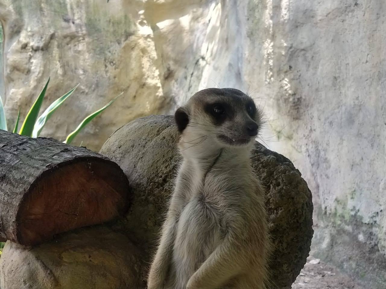
This article describes the case of a 5-year-old female meerkat born under human care at the Cali Zoo in Colombia, which presented a report of abnormal behavior. Upon clinical examination, multifocal and symmetrical bilateral scabbed alopecia was observed. An ultrasonographic evaluation was carried out determining the presence of ovarian cysts. As a therapeutic alternative, an ovariohysterectomy (OVH) was performed with an excisional biopsy of both ovaries, which were sent to the pathology laboratory. The histopathological findings concluded a neoplasm of the left ovary consistent with a Luteoma. Nonetheless, the right ovary did not present significant lesions. During the surgery, blood samples were taken which indicated that the patient presented regenerative anemia, blood chemistry with increased BUN and total proteins associated with hyperglobulinemia indicative of a chronic inflammatory process. The patient recovered satisfactorily from the surgery and after two weeks following the procedure, the dermatological lesions receded, as well as the behavioral problems previously reported.
Article visits 1150 | PDF visits
Downloads
- Jordan N, Do Linh San E. Suricata suricatta. The IUCN Red List of Threatened Species. 2015. https://dx.doi.org/10.2305/IUCN.UK.2015-4.RLTS.T41624A45209377
- Beke Graw, Marta Manser. Life history patterns and biology of the slender mongoose (Galerella sanguinea) in the Kalahari Desert. J Mammal. 2017; 98(2):332–338. https://doi.org/10.1093/jmammal/gyw178
- Maya-Pulgarin D, Gonzalez-Dominguez MS, Aranzazu-Taborda D, Mendoza N, Maldonado-Estrada JG. Histopathologic findings in uteri and ovaries collected from clinically healthy dogs at elective ovariohysterectomy: a cross-sectional study. J Vet Sci. 2017; 18(3):407-414. http://dx.doi.org/10.4142/jvs.2017.18.3.407
- Molly E. Church, Karen A. Terio, Keel MK. Procyonidae, Viverridae, Hyenidae, Herpestidae, Eupleridae, and Prionodontidae. Karen A. Terio, Denise Mc Aloose, Judy St. Leger. Pathology of Wildlife and Zoo Animals. Elsevier; 2018. https://doi.org/10.1016/B978-0-12-805306-5.00012-2
- Juan-Sallés C, Prats N, López S, Domingo M, Marco AJ, Morán JF. Epizootic disseminated toxoplasmosis in captive slender-tailed meerkats (Suricata suricatta). Vet Pathol. 1997; 34(1):1-7. http://dx.doi.org/10.1177/030098589703400101
- Bongiovann L, Di Girolamo N, Della Salda L, Massimi M, Romanucci M,Selleri P, Sudden death in a captive meerkat (suricata suricatta) with arterial medial and myocardial calcification, Asian Pacific Journal of Tropical Biomedicine. 2016; 6(4):357-359. http://dx.doi.org/10.1016/j.apjtb.2016.01.009
- Vladimir Jekl, Karel Hauptman. Reproductive medicine in ferrets.Vet Clin North Am Exot Anim Pract. 2017; 20(2):629-663. https://doi.org/10.1016/j.cvex.2016.11.016
- Victoria A.Reproductive system of the dog and cat Part 1 – the female system. Vet Nurs J. 2011; 26(2):43-45. http://dx.doi.org/10.1111/j.2045-0648.2010.00013.x
- Feldman and Nelson. Canine and feline endocrinology and reproduction. Second edition; Elsevier; 2014.
- Daniel Salazar C, Rosa Perales C. Diagnóstico Histopatológico de Neoplasias en Tracto Reproductivo de Caninos y Felinos Hembras Realizadas en el Laboratorio de Patología Animal de la Universidad Nacional Mayor de San Marcos (2007-2015). Rev Inv Vet Perú 2017; 28(2):468-475. http://dx.doi.org/10.15381/rivep.v28i2.13068
- Lourdes Caballero Flores. A propósito de un caso clínico: Tumor ovárico primario. Badajoz Veterinaria. 2017; 7:52-54. https://www.colegioveterinariosbadajoz.com/images/Revistas/2017/Numero7.pdf
- Corey FS, . Lawrence JA. Tumors of the Female Reproductive System. 5th edition. David Vail Small Animal Clinical Oncology. Elsevier; 2012. https://doi.org/10.1016/B978-1-4377-2362-5.00026-8
- Haimerl P, Arlt SP. Cystic ovaries and ovarian neoplasia in the female dog – a systematic review. Reprod Domest Anim. 2016; 51(1):3-11. https://doi.org/10.1111/rda.12781
- Darbaz I, Ergene O, Sonmez G, Aslan S. Ovarian Tumour in a Bitch: Diagnosis, Surgery and Recovery. Kafkas Univ Vet Fak Derg. 2017; 23(5):839-842 https://doi.org/10.9775/kvfd.2017.17718
- Knauf Y, Köhler K, Knauf S, Wehrend A. Histological classification of canine ovarian cyst types with reference to medical history. J Vet Sci. 2018; 19(6):725-734. https://doi.org/10.4142/jvs.2018.19.6.725
- Dalen WA, MacLachlan J. Tumors of the genital systems. In: Meuten DJ. Tumors in Domestic Animals. fifth edition. John Wiley & Sons; 2017. https://doi.org/10.1002/9781119181200.ch16
- Huynh M, Laloi F. Diagnosis of liver disease in domestic ferrets (Mustela putorius). Vet Clin North Am Exot Anim Pract. 2013; 16(1):121-144. http://dx.doi.org/10.1016/j.cvex.2012.10.003
- Yamini B, VanDenBrink PL, Refsal KR. Ovarian steroid cell tumor resembling luteoma associated with hyperadrenocorticism (Cushing’s disease) in a dog. Vet Pathol. 1997; 34(1):57-60. http://dx.doi.org/10.1177/030098589703400112
Information


