Gene expression analysis of bovine granulosa cells from growing follicles
Expresión génica en células de granulosa bovinas de folículos en crecimiento


This work is licensed under a Creative Commons Attribution-NonCommercial-ShareAlike 4.0 International License.
Show authors biography
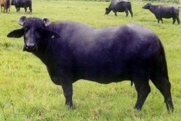
Objetive. This work compares granulosa cell gene expression using RNA analysis from pre-ovulatory follicles from two different bovine species (buffaloes and cattle). Materials and methods. The RNA was obtained from granulosa cells from ovaries of 10 buffaloes and cattle obtained at the local slaughterhouse, was sequenced with Novaseq, and the differential expression was analyzed using EdgeR in Bioconductor, and the function was assigned according to gene ontology terms. Results. Differential gene expression analyzes shown significant differences between species, but the most important feature is the low participation of genes associated with the reproductive process of follicular development, highlighting the importance of paracrine control of the ovary. It was found that between buffaloes and cattle, there is practically no correspondence in the gene expression of the physiological states evaluated; 6137 genes show differential expression between the two species. Conclusions. Each species has its way of performing the same process. The differences in the expression of the genes associated with oxidative phosphorylation are evident, and new ways to look at the presented results are required to understand the biological significance of the findings.
Article visits 682 | PDF visits
Downloads
- Buntjer JB, Otsen M, Nijman IJ, Kuiper MTR, Lenstra JA. Phylogeny of bovine species based on AFLP fingerprinting. Heredity. 2002; 88(1):46-51. https://doi.org/10.1038/sj.hdy.6800007.
- Gimenes LU, Ferraz ML, Fantinato-Neto P, Chiaratti MR, Mesquita LG, Sá Filho MF, Meirelles FV, Trinca LA, Rennó FP, Watanabe YF, Baruselli PS. The interval between the emergence of pharmacologically synchronized ovarian follicular waves and ovum pickup does not significantly affect in vitro embryo production in Bos indicus, Bos taurus, and Bubalus bubalis. Theriogenology. 2015; 83(3):385-93. https://doi.org/10.1016/j.theriogenology.2014.09.030
- Drost M. Bubaline versus bovine reproduction. Theriogenology. 2007; 68(3):447–449. https://doi.org/10.1016/j.theriogenology.2007.04.012
- Scaramuzzi RJ, Baird DT, Campbell BK, Driancourt MA, Dupont J, Fortune JE, Gilchrist RB, Martin GB, McNatty KP, McNeilly AS, Monget P. Regulation of folliculogenesis and the determination of ovulation rate in ruminants. Reprod. Fert. Dev. 2011; 23(3):444-467. https://doi.org/10.1071/RD09161.
- Gougeon A. Human ovarian follicular development: from activation of resting follicles to preovulatory maturation. Annal d’endocrin. 2010; 71(3):132–43. https://doi.org/10.1016/j.ando.2010.02.021
- Khan DR, Landry DA, Fournier É, Vigneault C, Blondin P, Sirard M-A. Transcriptome meta-analysis of three follicular compartments and its correlation with ovarian follicle maturity and oocyte developmental competence in cows. Physiol. Genom. 2016; 48(8):633–643. https://doi.org/10.1152/physiolgenomics.00050.2016
- Nivet A-L, Bunel A, Labrecque R, Belanger J, Vigneault C, Blondin P, et al. FSH withdrawal improves developmental competence of oocytes in the bovine model. Reproduction. 2012; 143(2):165–171. https://doi.org/10.1530/REP-11-0391
- Robert C. Microarray analysis of gene expression during early development: A cautionary overview. Reproduction. 2010; 140:787–801. https://doi.org/10.1530/REP-10-0191
- Li J, Liu J, Liu S, Plastow G, Zhang C, Wang Z, et al. Integrating RNA-seq and GWAS reveals novel genetic mutations for buffalo reproductive traits. Anim Reprod Sci. 2018; 197:290-295 https://doi.org/10.1016/j.anireprosci.2018.08.041
- Li J, Li Z, Liu S, Zia R, Liang A, Yang L. Transcriptome studies of granulosa cells at different stages of ovarian follicular development in buffalo. Anim Reprod Sci. 2017; 187:181–192. https://doi.org/10.1016/j.anireprosci.2017.11.004
- Daetwyler HD, Capitan A, Pausch H, Stothard P, Van Binsbergen R, Brøndum RF, Liao X, Djari A, Rodriguez SC, Grohs C, Esquerre D. Whole-genome sequencing of 234 bulls facilitates mapping of monogenic and complex traits in cattle. Nature Genetics. 2014; 46(8):858-865. https://doi.org/10.1038/ng.3034
- Hatzirodos N, Hummitzsch K, Irving-Rodgers HF, Harland ML, Morris SE, Rodgers RJ. Transcriptome profiling of granulosa cells from bovine ovarian follicles during atresia. BMC Genom. 2014; 15(1):1-26. https://doi.org/10.1186/1471-2164-15-40
- Rao JU, Shah KB, Puttaiah J, Rudraiah M. Gene Expression Profiling of Preovulatory Follicle in the Buffalo Cow: Effects of Increased IGF-I Concentration on Periovulatory Events. PLoS ONE. 201; 6(6):e20754 https://doi.org/10.1371/journal.pone.0020754.
- Marin DF, Souza EB, Brito VC, Nascimento CV, Ramos AS, Rolim ST, Costa NN, Cordeiro MD, Santos SD, Ohashi OM. In vitro embryo production in buffaloes: from the laboratory to the farm. Anim. Reprod. Br. 2019; 16:260-266. https://doi.org/10.21451/1984-3143-AR2018-0135
- Tremblay PG, Sirard M-A. Transcriptomic analysis of gene cascades involved in protein kinase A and C signaling in the KGN line of human ovarian granulosa tumor cells. Biol Reprod. 2017; 96(4):855–865. https://doi.org/10.1093/biolre/iox024
- Hummitzsch K, Irving-Rodgers HF, Hatzirodos N, Bonner W, Sabatier L, Reinhardt DP, Sado Y, Ninomiya Y, Wilhelm D, Rodgers RJ. A new model of development of the mammalian ovary and follicles. PloS One. 2013; 8(2):e55578 https://doi.org/10.1371/journal.pone.0055578
- Khan DR, Guillemette C, Sirard MA, Richard FJ. Characterization of FSH signalling networks in bovine cumulus cells: a perspective on oocyte competence acquisition. Mol. Hum. Reprod. 2015; 21(9):688–701. https://doi.org/10.1093/molehr/gav032
- McGettigan PA, Browne JA, Carrington SD, Crowe MA, Fair T, Forde N, et al. Fertility and genomics: comparison of gene expression in contrasting reproductive tissues of female cattle. Reprod Fert Dev. 2016; 28(2):11. https://doi.org/10.1071/RD15354
- Takeo S, Kawahara-Miki R, Goto H, Cao F, Kimura K, Monji Y, et al. Age-associated changes in gene expression and developmental competence of bovine oocytes, and a possible countermeasure against age-associated events. Mol Rep Dev. 2013; 80(7):508–521. https://doi.org/10.1002/mrd.22187
Information


