Computed tomography examination of the os cordis in a lamb (Ovis aries Linnaeus, 1758)
Tomografía computarizada del hueso cardíaco en un cordero (Ovis aries Linnaeus, 1758)


This work is licensed under a Creative Commons Attribution-NonCommercial-ShareAlike 4.0 International License.
Show authors biography
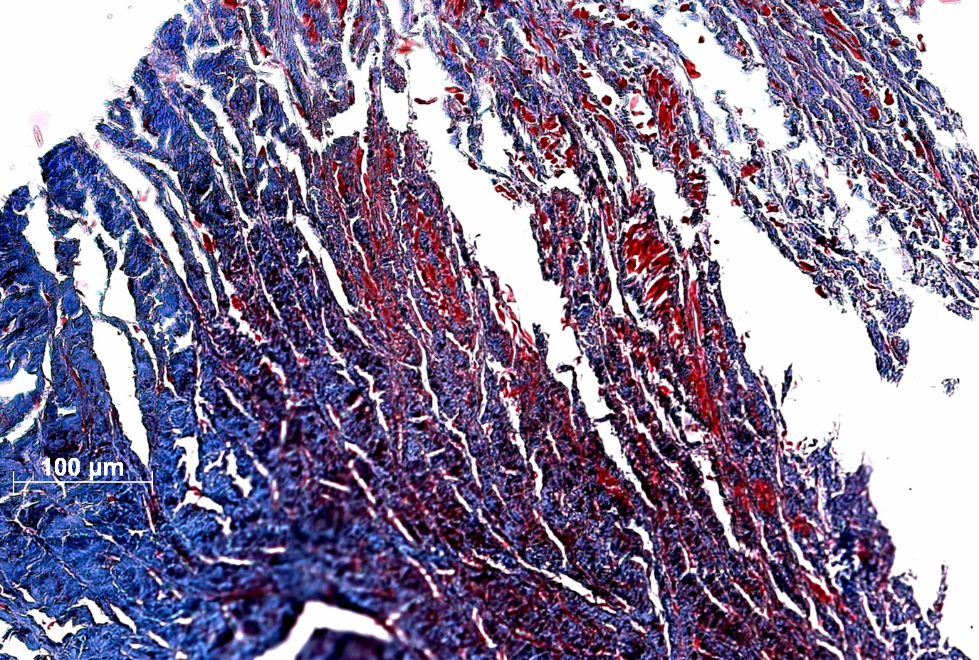
Objective. The present paper aims to investigate the role of computed tomography as an imaging technique of diagnosis in the identification of the os cordis in ovine, and also how this anatomical structure is morphotopographically characterized in macro and microscopic contexts, seeking to contribute for its functional understanding. Materials and method. The heart of a young male ovine had been donated to the Laboratory of Animal Anatomy of the Surgery Department of the FMVZ/USP, first being subject to a post-mortem examination by means of the cardiac “shedding” (transverse cross sectioning of the heart). A tomographic examination of the anatomic specimen was carried out, as well as the dissection and histological analysis of the collected sample. Results. The results indicate the presence of an osseous structure of 6.39 mm in length, located in the aortic valve of the heart, next to a ring comprised by three valve flaps, tendinous cords, and papillary muscles. The histological findings consist of fibrous connective tissue, cancellous bone tissue, and calcified hyaline cartilage wherein the cardiomyocytes are attached. Conclusions. It is concluded that computed tomography, even though seldom applied to animals of zootechnical interest, poses as an effective tool for the visualization of the os cordis in lambs.
Article visits 1151 | PDF visits
Downloads
- Mohapatra A, Shinde AK, Singh R. Sheep milk: A pertinent functional food. Small Ruminant Res. 2019; 181:6-11. https://doi.org/10.1016/j.smallrumres.2019.10.002
- Hermuche PM, Maranhão RL, Guimarães RF, De Carvalho OA, Gomes RA, Paiva SR, McManus C. Dynamics of sheep production in Brazil. ISPRS Int J Geo-Inf. 2013; 2(3):665-679. https://doi.org/10.3390/ijgi2030665
- Butters TD, Aslanidi OV, Zhao J, Smaill B, Zhang H. A novel computational sheep atria model for the study of atrial fibrillation. Interface Focus. 2013; 3(2):20120067. https://doi.org/10.1098/rsfs.2012.0067
- Rienzo M, Imbault J, El Boustani Y, Beurton A, Sampedrano CC, Pasdois P, et al. A total closed chest sheep model of cardiogenic shock by percutaneous intracoronary ethanol injection. Sci Rep. 2020; 10(1):1-3. https://doi.org/10.1038/s41598-020-68571-5
- Monreal G, Sherwood LC, Sobieski MA, Giridharan GA, Slaughter MS, Koenig SC. Large animal models for left ventricular assist device research and development. Asaio J. 2014; 60(1):2-8. https://doi.org/10.1097/MAT.0000000000000005
- Katz MG, Kendle AP, Fargnoli AS, Mihalko KL, Bridges CR. Sheep (Ovis aries) as a model for cardiovascular surgery and management before, during, and after cardiopulmonary bypass. J Am Assoc Lab Anim Sci. 2015; 54(1):7. https://www.ncbi.nlm.nih.gov/pmc/articles/PMC4311734
- Saunders JD, O’Malley CD. Andreas Vesalius de Bruxelas. De humani Corporis Fabrica. Epitome. Tabulae Sex. Unicamp; 2002.
- Mohammadpour A, Arabi M. Morphological study of the heart and os cordis in sheep and goat. Indian Vet J. 2007;84(3):284-287. https://profdoc.um.ac.ir/paper-abstract-103021.html
- International Committee on Veterinary Gross Anatomical Nomenclature. Nomina Anatomica Veterinaria. 6th ed. Hannover: Editorial Committee; 2017. http://www.wava-amav.org/wava-documents.html
- Nute JL, Le Roux L, Chandler AG, Baladandayuthapani V, Schellingerhout D, Cody DD. Differentiation of low-attenuation intracranial hemorrhage and calcification using dual-energy computed tomography in a phantom system. Invest Radiol. 2015; 50(1):9. https://doi.org/10.1097/RLI.0000000000000089
- Frink RJ, Merrick B. The sheep heart: Coronary and conduction system anatomy with special reference to the presence of an os cordis. Anat Rec. 1974; 179(2):189-199. https://doi.org/10.1002/ar.1091790204
- Abolhasanzadeh A, Mastary Farahani R, Alexanian S. Histological study of os cordis of sheep. Feyz. 2000; 3(4):25-29 http://feyz.kaums.ac.ir/article-1-476-en.html
- König HE, Liebich HG, editors. Veterinary anatomy of domestic animals: Textbook and colour atlas. Georg Thieme Verlag; 2020.
- Akhtar S, Hossain FM, Siddiqui MS, Alam M, Islam MN. Morphology and Morphometrical studies on Semi lunar Heart valves of Indigenous Cattle (Bos indicus). Int J Nat Sci. 2011; 1(1):7-11. https://doi.org/10.3329/ijns.v1i1.8608
- Ghonimi W, Balah A, Bareedy MH, Abuel-Atta AA. Os cordis of the mature dromedary camel heart (Camelus dromedaries) with special emphasis to the cartilago cordis. J Veterinar Sci Technolo. 2014; 5(4):1. https://doi.org/10.4172/2157-7579.1000193
- Moittié S, Baiker K, Strong V, Cousins E, White K, Liptovszky M, Redrobe S, Alibhai A, Sturrock CJ, Rutland CS. Discovery of os cordis in the cardiac skeleton of chimpanzees (Pan troglodytes). Sci Rep. 2020; 10(1):1-1. https://doi.org/10.1038/s41598-020-66345-7
- Almeida MC, Sánchez-Quintana D, Davis N, Charles FR, Chikweto A, Sylvester W, Loukas M, Anderson RH. The ox atrioventricular conduction axis compared to human in relation to the original investigation of Sunao Tawara. Clin Anat. 2020; 33(3):383-393. https://doi.org/10.1002/ca.23524
- Cardoso PGS, Pinto MPR, Moroz LR, Fontes TN, Santos RS, Freitas JL, Nogueira VA, Peixoto TC. Dystrophic mineralization in uremic dogs: na uptade. Pesq Vet Bras. 2019; 39(11):889-899. https://doi.org/10.1590/1678-5150-pvb-6250
- Schwarz T, Sullivan M, Störk CK, Willis R, Harley R, Mellor DJ. Aortic and cardiac mineralization in the dog. Vet Radiol Ultrasoun. 2002; 43(5):419-27. https://doi.org/10.1111/j.1740-8261.2002.tb01028.x
- Galas A, Hryniewiecki T, Michałowska I, Kępka C, Abramczuk E, Orłowska-Baranowska E, Rużyłło W. Aortic valve calcification in 499 consecutive patients referred for computed tomography.
- Arch Med Sci 2015; 11(5):952-957. https://doi.org/10.5114/aoms.2015.47874
- Otsuka F, Sakakura K, Yahagi K, Joner M, Virmani R. Has our understanding of calcification in human coronary atherosclerosis progressed? Arterioscler Thromb Vasc Biol. 2014; 34(4):724-736. https://doi.org/10.1161/ATVBAHA.113.302642
- Owens DS, Budoff MJ, Katz R, Takasu J, Shavelle DM, Carr JJ, Heckbert SR, Otto CM, Probstfield JL, Kronmal RA, O’Brien KDO. Aortic valve calcium independently predicts coronary and cardiovascular events in a primary prevention population. JACC: Cardiovasc Imaging. 2012; 5(6):619-625. https://doi.org/10.1016/j.jcmg.2011.12.023
- Sato Y, Jinnouchi H, Sakamoto A, Cornelissen A, Mori M, Kawakami R, Kawai K, Virmani R, Finn AV. Calcification in human vessels and valves: from pathological point of view. AIMS Mol Sci. 2020; 7(3):183-210. https://doi.org/10.3934/molsci.2020009
- Zheng KH, Tsimikas S, Pawade T, Kroon J, Jenkins WS, Doris MK, White AC, Timmers NK, Hjortnaes J, Rogers MA, Aikawa E. Lipoprotein (a) and oxidized phospholipids promote valve calcification in patients with aortic stenosis. J Am Coll Cardiol. 2019; 73(17):2150-2162. https://doi.org/10.1016/j.jacc.2019.01.070
- Mantovani A, Pernigo M, Bergamini C, Bonapace S, Lipari P, Valbusa F, Bertolini L, Zenari L, Pichiri I, Dauriz M, Zoppini G. Heart valve calcification in patients with type 2 diabetes and nonalcoholic fatty liver disease. Metabolism. 2015; 64(8):879-887. https://doi.org/10.1016/j.metabol.2015.04.003
- Sakamoto A, Guo L, Virmani R, Finn AV. Is there a role for activated platelets in progression of aortic valve calcification? Eur Heart J. 2019; 40(17):1374–1377. https://doi.org/10.1093/eurheartj/ehy696
- Carrai P, Camarri S, Pondrelli CR, Gonnelli S, Caffarelli C. Calcification of cardiac valves in metabolic bone disease: an updated review of clinical studies. Clin Interv Aging. 2020;15:1085-1095. https://doi.org/10.2147/CIA.S244063
- Keywords: Monogastric livestock; nitrogen content; physicochemical properties; extrusion; standardization
Information


