Atypical eosinophilic granuloma in a dog. Case report
Granuloma eosinofílico atípico en un perro. Reporte de caso


This work is licensed under a Creative Commons Attribution-NonCommercial-ShareAlike 4.0 International License.
Show authors biography
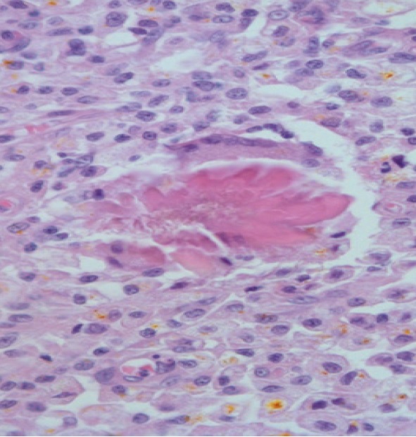
The eosinophilic granuloma is characterized by dense cellular infiltration of eosinophils, macrophages, and mast cells, in addition to collagenolysis foci. Canine eosinophilic granuloma is a rare inflammatory skin disease that affects the oral mucosa. However, the cutaneous form is more evident and can be in the scrotum, testicles, perineum, and flanks. The etiology of eosinophilic granuloma in dogs is not clear but allergic processes can originate granulomatous nodules or plaques. Affected dogs may experience itching, swelling, and, occasionally, pain. The histopathological analysis is necessary for definitive diagnosis. The present case report described a 12-year-old male dog, Poodle, with a history of swelling, itching, and pain in the scrotum. Complete clinical staging was performed through blood count, biochemistry analysis, thoracic radiographs, abdominal ultrasound, and electrocardiogram. The dog was submitted to surgical removal followed by histopathologic evaluation. Eosinophilic granuloma should be included in the differential diagnosis of solitary masses in the scrotal region.
Article visits 2070 | PDF visits
Downloads
- Hnilica KA, Patterson AP. Editores. Dermatology of small animals: colorful atlas and therapeutic guide. 4a ed. Missouri: W.B. Saunders Company; 2018. https://doi.org/10.1016/C2014-0-01191-4
- Lommer MJ. Oral inflammation in small animals. Vet Clin North Am Small Anim Pract. 2013; 43(3):555-571. https://doi.org/10.1016/j.cvsm.2013.02.004
- Kim JH, Jung JY, Kang SC, Lee YR, Lee JY, Hwang EK et al. Eosinophilic granulomas in two dogs. Korean J Vet Res. 2011; 51(1):61-64. https://doi.org/10.14405/kjvr.2011.51.1.069
- Declercq J, Vercauteren, G. Necrotizing eosinophilic dermatitis in three dogs. Vlaams Diergen Tijds. 2019; 88(2):91-96. https://doi.org/10.21825/vdt.v88i2.16030
- Weiss DJ & Wardrop, Schalm’s KJ. Editores. Schalm’s Veterinary Hematology. 6ª ed. Iowa: Blackwell Publishing; 2011.
- Vercelli A, Cornegliani L, Portigliotti L. Eyelid eosinophilic granuloma in a Siberian husky. J Small Anim Pract. 2005; 46(1):31–33. https://doi.org/10.1111/j.1748-5827.2005.tb00272.x
- Stockham SL, Scott MA. Editores. Fundamentals of Veterinary Clinical Pathology. 2a ed. Iowa: Blackwell Publishing; 2013.
- Knight EC, Shipstone MA. Canine eosinophilic granuloma of the digits treated with prednisolone and chlorambucil. Vet Dermatol 2016; 27(5):446–119. https://doi.org/10.1111/vde.12355
- Tellado MN, Michinski SD, Olaiz N, Maglietti F, Marshall Guillermo. Canine Oral Eosinophilic Granuloma Treated with Electrochemotherapy. Case Rep Vet Med. 2014; 2014(1):1–5. https://doi.org/10.1155/2014/519197
- Mali B, Jarm T, Snoj M, Sersa G, Miklavcic D. Antitumor effectiveness of electrochemotherapy: A systematic review and meta-analysis. Eur J Surg Oncol. 2013; 39(1):4–16. https://doi.org/10.1016/j.ejso.2012.08.016
- Kotnik T, Kramar P, Pucihar G, Miklavčič D, and M. Tarek. Cell membrane electroporation—part 1: the phenomenon. IEEE Electric Insul M. 2012; 28(5):14–23. https://doi.org/10.1109/MEI.2012.6268438
Information


