Show authors biography
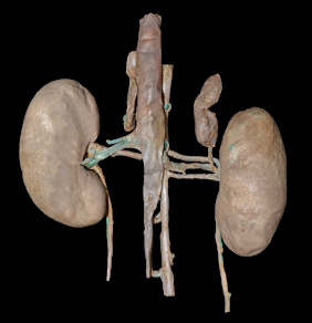
Objective. The aim of this study was explored the duplicity of renal artery in a specimen of Cerdocyon thous, focusing on the possibilities of clinical-surgical implication of this anatomical variation. Materials and Methods. Were dissected 32 specimens of Cerdocyon thous, obtained from the collections of the Laboratório de Ensino e Pesquisa em Morfologia dos Animais Domésticos e Selvagens do Departamento de Anatomia Animal e Humana, da Universidade Federal Rural do Rio de Janeiro e Laboratório de Anatomia Animal da Universidade Federal do Pampa. Results. Were observed a numerical variation in the left renal artery in an adult female cadaver. The left kidney had two renal arteries, one cranial and another caudal. The first renal artery of the left kidney, measuring 2.25 cm in length, originated laterally from the abdominal aorta at the level of the third lumbar vertebra. Moreover, it emanated two pre-hilar branches, one dorsal and one ventral, with the ventral branch supplying also to the adrenal gland. The second renal artery also originated laterally from the abdominal aorta at the level of the third lumbar vertebra and measured 2.36 cm in length. It also emitted two pre-hilar branches, one cranial and another caudal, which emitted the ureteral branch. Conclusions. Numerical variations of the renal arteries should be considered in the execution of surgical, radiological and experimental procedures in order to avoid mistakes made due to lack of knowledge of the possibility these variations both in domestic and wild animals.
Article visits 1103 | PDF visits
Downloads
- Machado FA, Hingst-Zaher E. Investigating South American biogeographic history using patterns of skull shape variation on Cerdocyon thous (Mammalia: Canidae). Biol J Linn Soc. 2009; 98(1):77-84. https://doi.org/10.1111/j.1095-8312.2009.01274.x
- Perini FA, Russo CAM, Schrago CG. The evolution of South American endemic canids: a history of rapid diversification and morphological parallelism. J Evol Biol. 2010; 23(2):311-322. https://doi.org/10.1111/j.1420-9101.2009.01901.x
- Trigo TC, Rodrigues MLF, Kasper CB. Carnívoros Continentais. In: Weber MM, Roman C, Cáceres NC. Mamíferos do Rio Grande do Sul. Santa Maria: UFSM; 2013. https://editoraufsm.com.br/mamiferos-do-rio-grande-do-sul
- Kasper CB, Trinca CS, Sanfelice D, Mazi FD, Trigo TC. Os Carnívoros. In: Gonçalves GL, Quintela FM, Freitas TRO. (eds.) Mamíferos do Rio Grande do Sul. Porto Alegre: Pacartes; 2014. https://www.catarse.me/mamiferosrs
- Sampaio FJ, Aragão AH. Anatomical relationship between the intrarenal arteries and the kidney collecting system. J Urol 1990; 143(4):679-681. https://doi.org/10.1016/s0022-5347(17)40056-5
- Sampaio FJB, Passos MARF. Renal arteries: anatomic study for surgical and radiological practice. Surg Radiol Anat 1992; 14(2):113-117. https://link.springer.com/article/10.1007/BF01794885
- König HE, Liebich HG. Anatomia dos animais domésticos: texto e atlas colorido. 6th ed. Porto Alegre: Artmed; 2016.
- Reis RH, Tepe P. Variation in the pattern of renal vessels and their relation to the type of posterior vena vava in the dog (Canis familiaris). Am. J. Anat 1956; 99:1-15. https://doi.org/10.1002/aja.1000990102
- Sajjarengpong K, Adirektaworn A. The variations and patterns of renal arteries in dogs. The Thai Journal of Veterinary Medicine 2006; 36(1):39-46. www.tci-thaijo.org/index.php/tjvm/article/view/36332
- Abidu-Figueiredo M, Roza MS, Passos NC, Silva BX, Scherer PO. Artéria renal com dupla origem na porção abdominal da aorta em caprino. Acta Veterinaria Brasilica 2009; 3(1):38-42. https://doi.org/10.21708/avb.2009.3.1.970
- Almeida BB, Barreto UH, Costa OM, Abidu-Figueiredo M. Double renal artery in rabbits. Biosci J 2013; 29(5):1294-1295. http://www.seer.ufu.br/index.php/biosciencejournal/article/view/22287
- Machado LC, Roballo KCS, Cury FS, Ambrósio CE. Female repro-ductive system morphology of crab-eating fox (Cerdocyon thous) and cryopreservation of genetic material for animal germplasm bank en-richment. Anat Histol Embryol 2017; 46(6): 539-546. http://dx.doi.org/10.1111/ahe.12306
- Pestana FM, Roza MS, Hernandez JMF, Silva BX, Abidu-Figueiredo M. Artéria renal dupla em gato: relato de caso. Semina: Cien Agrar 2011; 32(1):325-330. http://dx.doi.org/10.5433/1679-0359.2011v32n1p327
- Khamanarong K, Prachaney P, Utraravichien A, Tong-Un T, Sri-paoraya K. Anatomy of renal arterial supply. Clin Anat 2004; 17:334-336. https://doi.org/10.1002/ca.10236
- Evans, H. E., A. Delahunta. Miller’s Anatomy of the Dog. 4th ed. St Louis (MO): Saunders Elsevier; 2013. https://www.elsevier.com/books/millers-anatomy-of-the-dog/evans/978-1-4377-0812-7
- Tavares DS, Varjão COV, Santos AH, Iamagute LS, Conceição AM, Barros SLB. Amputação de membro pélvico de cachorro-do-mato (Cerdocyon thous) devido à osteomielite pós cirurgia de correção de fratura: relato de caso. Rev Educ Cont Vet Med Zootec 2013; 11(3):98. https://www.revistamvez- crmvsp.com.br/index.php/recmvz/article/view/21397
- Sposito GC, Gorios A, Camargo LP, Campos MAR, Estrella JPN, Credie LFGA, et al. Anestesia epidural sacrococcígea em osteossíntese femoral em cachorro-do-mato (Cerdocyon thous). Relato de caso. Rev Educ Cont Vet Med Zootec 2016; 14(2):46. https://www.revistamvez-crmvsp.com.br/index.php/recmvz/article/view/31823
- Silva ASL, Feliciano MAR, Motheo TF, Oliveira JP, Kawanami AE, Werther K, et al. Mode B ultrasonography and abdominal Doppler in crab-eating-foxes (Cerdocyon thous). Pesq Vet Bras 2014; 34(Suppl 1):23-28. https://doi.org/10.1590/S0100-736X2014001300005
- Silva TR, Nogueira AFS, Santana AE. Avaliação dos perfis bio-químicos hepático e renal em Cerdocyon thous (Cachorro do mato) Sororreagentes a Leptospira spp. no território brasileiro. Ars Veteri-naria 2013; 29(4):9. http://dx.doi.org/10.15361/2175-0106.2013v29n4p9
- Piccoli RJ, Thomazoni D, Druziani JT, Hamamura M, Carvalho AL. Nefrectomia total unilateral em cachorro-do-mato (Cerdocyon thous). Acta Sci Vet 2017; 45(Suppl 1):228. http://www.ufrgs.br/actavet/45-suple-1/CR_228.pdf


